Schilddrüsenblutung

Endovascular
treatment of massive hemorrhage arising from inferior thyroid artery after fine needle aspiration of thyroid: a case report. a An axial neck CT take after thyroid FNA underwent on emergency department reveals a large hematoma (red asterisk) in the anterior neck space with anterior tracheal deviation and nodule in the right thyroid lobe with intra- and extra nodule air-bubbles caused by the fine needle aspiration. b Sagittal CT scan shows extravasation of contrast media suggesting active bleeding (red arrow) within the hematoma

Endovascular
treatment of massive hemorrhage arising from inferior thyroid artery after fine needle aspiration of thyroid: a case report. a Right subclavian angiography shows active bleeding (red arrow) in the neck, corresponding to the CT image. b Right thyrocervical trunk is selected and the culprit branch is identified as the right inferior thyroid artery (red arrow head)

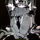
A Case of
Diffuse Thyroid Hematoma after Ultrasound-Guided Fine Needle Aspiration: Enhanced neck computed tomography. (A) The axial view shows diffuse swelling and heterogenous enhancement of both thyroid lobes. (B) The sagittal view shows diffuse non-enhancing soft-tissue density lesions of the retropharyngeal space and superior mediastinum.

Thyroid gland
hemorrhage after blunt neck trauma: case report and review of the literature. Contrast-enhanced computed tomography revealed a laceration of the right thyroid lobe with diffuse hematoma

Sudden
massive neck swelling due to hemorrhage of a thyroid adenoma: a case report. Axial computed tomography (CT) image showing the displacement of the larynx to the right and the inhomogeneous mass on the left.

Sudden
massive neck swelling due to hemorrhage of a thyroid adenoma: a case report. Sagittal computed tomography (CT) image showing the extent of the mass and the contrast enhancement in its caudal region.
Sudden
massive neck swelling due to hemorrhage of a thyroid adenoma: a case report. Coronal computed tomography (CT) image showing the possible connection of the mass to the thyroid gland.

Thyroid gland
rupture caused by blunt trauma to the neck. Computed tomography of the neck and chest. Computed tomography showing thyroid rupture, with a large hematoma (arrow) extending from the neck (a) to the mediastinum (b)


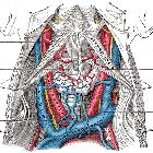
 Assoziationen und Differentialdiagnosen zu Schilddrüsenblutung: