Osteochondrosis intervertebralis

Intervertebral
osteochondrosis • Intervertebral osteochondrosis - Ganzer Fall bei Radiopaedia


Spinal
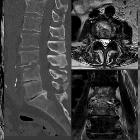
disorders mimicking infection. 68-year-old male with increasing back pain referred for pathologic confirmation and treatment of spondylodiscitis. Sagittal XR (a), Sagittal T1 (b), T2 (c), STIR (d) and T1 Fatsat after gadolinium R1 point 5 (e) confirm edema-type enhancing marrow signal abnormalities as well as disc hyperintensity (arrow), and narrowed L3–L4 space without disc enhancement. Following sagittal CT (f) R1 point 6 confirms the absence of endplates destruction and that the hypersignal on the underlying disc (L4–L5) corresponds to the vacuum phenomenon (star), biopsy performed under CT guidance (g) was negative. At 3 months, MRI control on sagittal T1 (h), STIR (i) and after contrast (j) shows stability of disco-vertebral features (arrow)
Intervertebral osteochondrosis represents the pathologic degenerative process involving the intervertebral disc and the respective vertebral body endplates (not necessarily symptomatic). It is believed to be different and a further stage of spondylosis deformans, which is a consequence of normal aging.
Terminology
It should not be confused with the general use of "osteochondrosis", which is a generic term given to a group of disorders that affect the progress of bone growth by bone necrosis.
Radiographic features
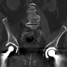
It is characterized by disc space narrowing, severe disc fissuring, vacuum phenomenon, endplate cartilage erosions, and vertebral body reactive changes (osteophytes and Modic changes).
Siehe auch:
und weiter:

 Assoziationen und Differentialdiagnosen zu Osteochondrosis intervertebralis:

