Unterscheidung infektiöse vs. nicht infektiöse Discitis Spondylodiscitis

Spinal
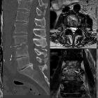
disorders mimicking infection. 32-year-old male admitted for exacerbation of back pain and inflammatory markers. T1 (a), T2WI (b), and STIR- R1 point 7 (c) show inflammatory endplate changes at the L3–L4 level mimicking a Schmorl node (arrow). Note that nuclear cleft sign is preserved (star). Enhancement in T1 fat-suppressed sequence after contrast (d) without disc enhancement or inflammatory paraspinal tissue. The same pattern is seen in the D11–D12 disc space (long arrow), associated with Romanus feature R1 point 8 at L5 body margin (short arrow)

Spinal
disorders mimicking infection. 70-year-old male with chronic history of back pain, recently exacerbated, and inflammatory laboratory markers. Sagittal T1WI (a) shows hypointense discal changes in the L4–L5 space and epidural space (arrow). Bony erosion of endplates is also seen (black arrow) at the L5 level (b, c). Sagittal T2WI (b) shows hyperintense areas in the L4–L5 disc space (star). Contrast-enhanced sagittal and axial T1WI (c, d) show enhancement of disc space, epidural space, left facet joint, and paraspinal muscles (arrow). Non-enhancing foci correspond to crystal material (arrow). Axial and sagittal slices of CT (e, f) show in L4–L5, disc space erosions, sclerotic vertebral bodies, and confirm gout tophi in muscles (short arrow)

Spinal
disorders mimicking infection. 72-year old male with amyotrophic lateral sclerosis presenting with subacute back pain, fever and inflammation in laboratory findings. Sagittal T2 (a), T1 (b) WI, DWI (c) and T1 after contrast on sagittal (d) and axial planes (e) show multiple contiguous erosive disco-vertebral disease on dorsal spine with local deformity and advanced destruction (arrow). No diffusion restriction was noticed on DWI (black arrow). Note anterior and posterior involvement (star). Reformatted sagittal CT (f) shows bone sclerosis and subtle vacuum phenomena in the disc (arrow). R1 Point 11The final diagnosis was Charcot spine

Spinal
disorders mimicking infection. 47-year-old male presenting with inflammatory back pain. Sagittal T1 (a) and R1 point 12 T2 (b)-WI show erosive L4-L5 endplates (arrow) associated on Sagittal STIR sequence (c) to a disc hypersignal (arrow) and on sagittal (d) and axial (e) T1 Fat Sat WI enhanced sequences to an enhancement of vertebral bodies and surrounding tissues as epidural fat space without abscess (star). Axial CT (f) on lumbar spine shows erosive pattern of vertebra (arrow) and Oblique reformatted CT (g) of anterior chest shows involvement with sclerotic lesions typical of SAPHO disease (star)

Spinal
disorders mimicking infection. 68-year-old male with increasing back pain referred for pathologic confirmation and treatment of spondylodiscitis. Sagittal XR (a), Sagittal T1 (b), T2 (c), STIR (d) and T1 Fatsat after gadolinium R1 point 5 (e) confirm edema-type enhancing marrow signal abnormalities as well as disc hyperintensity (arrow), and narrowed L3–L4 space without disc enhancement. Following sagittal CT (f) R1 point 6 confirms the absence of endplates destruction and that the hypersignal on the underlying disc (L4–L5) corresponds to the vacuum phenomenon (star), biopsy performed under CT guidance (g) was negative. At 3 months, MRI control on sagittal T1 (h), STIR (i) and after contrast (j) shows stability of disco-vertebral features (arrow)

Spinal
disorders mimicking infection. R1 point 9: 78-year-old male with sepsis and thoracic back pain with ankylosing spondylitis. MRI (sagittal T1 (a), T2 (b), STIR (c) and T1 Fat Sat Gadolinium (d)) shows T6-T7 disc hypersignal, enhanced after administration of contrast (arrow) and marked inflammatory surrounding tissue in axial T2 (f) and axial T1FS with contrast (g) (star). Note the elevated medullary signal intensity secondary to compression (black arrow). Reformatted CT (e) shows ankylosed spine and transdiscal fracture (star). Discal biopsy (h) was performed to exclude an infection R1 point 10. Andersson discitis was diagnosed
Unterscheidung infektiöse vs. nicht infektiöse Discitis Spondylodiscitis
Siehe auch:

 Assoziationen und Differentialdiagnosen zu Unterscheidung infektiöse vs. nicht infektiöse Discitis Spondylodiscitis:


