trapezoid


The trapezoid bone (also known as the os trapezoideum or the lesser multangular) is the smallest carpal bone in the distal row, sitting lateral to the capitate. The trapezium and trapezoid are collectively known as the multangulars.
Gross anatomy
Osteology
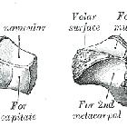
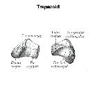
The trapezoid is an irregular, boot-shaped bone. The dorsal surface is larger than the palmar surface and is elongated in the transverse direction. It has a smaller square-shaped palmar surface projecting out of the larger dorsal portion. The palmar surface connects to the dorsal portion slightly laterally. The distal surface is triangular, with a palmar apex. The distal articulation surface has two small concave facet surfaces medially and laterally, with an overall convex appearance. The medial and lateral surfaces are both narrow, with the medial surface concave and the lateral surface convex in appearance.
Articulations
The trapezoid articulates with the scaphoid, capitate, trapezium and the base of the second metacarpal.
- proximal surface: scaphoid, comprising part of the triscaphe joint
- distal surface: base of the second metacarpal
- lateral surface: trapezium
- medial surface: capitate
Attachments
Musculotendinous
- origin of deep head of flexor pollicis brevis from palmar surface
- origin of adductor pollicis (oblique head) from distal ulnar palmar surface (variably)
The dorsal surface has no muscle attachments.
Ligamentous
- trapeziotrapezoid
- trapeziocapitate
- dorsal and volar carpometacarpal
- dorsal intercarpal
- scaphotrapezium-trapezoid
Arterial supply
The dorsal intercarpal and basal metacarpal arches, as well as the radial recurrent artery, provide the vascularity of the trapezoid. Vessels enter through both the central dorsal and palmar surfaces. The dorsal vessels provide the primary vascularity, the dorsal 70% of the bone. There are no anastomoses between the dorsal and palmar surface entering vessels.
Variant anatomy
Accessory bones associated with the trapezoid may be mistakenly viewed as fractures. See Accessory ossicles of the wrist.
The trapezoid may occasionally have an attachment from the origin of the oblique head of the adductor pollicis.
Development
Ossification
Trapezoid ossification begins around the fourth year in females and approximately twelve months later in males. It generally has a single ossification center.
Related investigations
Plain radiograph
The trapezoid may be visualized on a number of series of the distal upper limb including:
Cross-sectional imaging
CT or MRI imaging will demonstrate the trapezoid and should be considered if there is clinical suspicion of occult injury.
Related pathology
Plain radiograph
Like the capitate, the trapezoid has a protected position and is the least commonly fractured carpal bone. Trapezoid fractures usually occur at the dorsal rim or body. An isolated trapezoid fracture is rare; it is often associated with a second metacarpal fracture. A trapezoid fracture may be difficult to visualize on routine radiograph imaging and may be assisted by oblique views.
Dorsal dislocations and less commonly palmar dislocations can be associated with capsular ligamentous rupture. Such injuries are often the result of axial loading of the index metacarpal.
Siehe auch:
und weiter:

 Assoziationen und Differentialdiagnosen zu Os trapezoideum:

