Pilozytisches Astrozytom des Kleinhirns

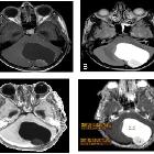
Cerebellar
pilocytic astrocytoma: MR spectroscopy. Shows cystic lesion with eccentric enhancing mural nodule in posterior fossa

Cerebellar
pilocytic astrocytoma: MR spectroscopy. T1WI shows hypointense lesion in the posterior fossa

Cerebellar
pilocytic astrocytoma: MR spectroscopy. T2WI shows hyperintense lesion in posterior fossa with effaced 4th ventricle and triventricular hydrocephalus with eccenteric intermediate signal intensity solid component.

Cerebellar
pilocytic astrocytoma: MR spectroscopy. Post contrast T1WI shows peripheral enhancing cystic lesion with eccentric mural nodule.

Cerebellar
pilocytic astrocytoma: MR spectroscopy. MRS shows laclate doublet at 1.3ppm and elevated CHO peak at 3.2ppm and reduced NAA peak and increase cho/Cr and cho/NAA ratio.
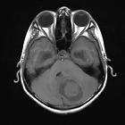
Pilocytic
astrocytoma - MRI appearance. MRI T1W axial image shows a well-defined hypointense lesion with inner hypo-isointense area in left cerebellum.

Pilocytic
astrocytoma - MRI appearance. MRI T2W axial image shows a well-defined hyperintense lesion with inner hypointense area noted in left cerebellum. The lesion shows mild perilesional oedema and causes compression over 4th ventricle resulting in mild hydrocephalus.

Pilozytisches Astrozytom des Kleinhirns
Siehe auch:
und weiter:


 Assoziationen und Differentialdiagnosen zu Pilozytisches Astrozytom des Kleinhirns: