zerebelläres Astrozytom


zerebelläres Astrozytom
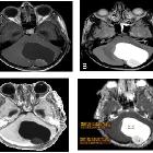
Astrozytom Radiopaedia • CC-by-nc-sa 3.0 • de
Astrocytic tumors are primary central nervous system tumors that either arises from astrocytes or appear similar to astrocytes on histology having arisen from precursor cells. They are the most common tumors arising from glial cells.
They can be divided into those that are diffuse in growth (the vast majority, generally having higher grade and poorer prognosis) and those that are localized (tend to have a lower grade and better prognosis).
- diffuse
- diffuse astrocytoma: WHO grade II (10-15% of astrocytomas)
- anaplastic astrocytoma: WHO grade III (25% of astrocytomas)
- glioblastoma: WHO grade IV (50-60% of astrocytomas)
- localized
- pilocytic astrocytoma: WHO grade I
- subependymal giant cell astrocytoma: WHO grade I
- pilomyxoid astrocytoma: ~WHO grade II
- pleomorphic xanthoastrocytoma: WHO grade II
Additionally, some tumors also contain astrocytic components, and it is often this component that dictated biological behavior. Examples include:
All these tumors have strikingly differing imaging appearances, treatment and prognosis and thus they are, along with spinal astrocytomas discussed separately.
NB: gliomatosis cerebri, fibrillary astrocytoma and protoplasmic astrocytoma are no longer recognized in the current WHO classification of CNS tumors
Siehe auch:


 Assoziationen und Differentialdiagnosen zu zerebelläres Astrozytom: