glomus tympanicum


Glomus tympanicum paragangliomas (chemodectomas) are the most common middle ear tumour.
Epidemiology
There is a female predominance (M: F = 1:3); presentation is most common when patients are more than 40 years old .
Clinical presentation
May be incidental but symptomatic masses produce pulsatile tinnitus, otalgia, or conductive hearing loss .
Pathology
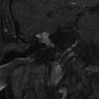
Glomus tympanicum paragangliomas arise from the Jacobson nerve at the cochlear promontory.
Radiographic features
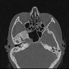
CT is usually the best modality to assess these masses.
CT
- soft tissue mass lateral to the cochlear promontory
- if large may fill the middle ear cavity, and invade the Eustachian tube or mastoid
- ossicles may or not be destroyed and may simply be encased
- surrounding bony destruction may be present in aggressive tumours
- intact jugular bulb
The Glasscock-Jackson and Fisch classifications of glomus tumours are based on the local extension of the tumour and their effect on mortality and morbidity. Glomus tympanicum paragangliomas are considered type A tumour, as they are limited to the middle ear cavity.
Treatment and prognosis
Surgical resection is the treatment of choice .
Differential diagnosis
On imaging consider:
- glomus jugulare paraganglioma
- permeative destruction of the floor of the middle ear
- involving the jugular foramen
- presents with a dehiscent jugular bulb
- facial nerve schwannoma
- pedunculated mass arising from the facial nerve
- involving the tympanic segment of the facial nerve
- congenital cholesteatoma
- no enhancement on post-contrast T1 MRI
See also
Siehe auch:
und weiter:


 Assoziationen und Differentialdiagnosen zu glomus tympanicum tumour:
