scrotal calculus
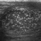

A rare case
of extratesticular scrotal epidermoid cyst. A non-vascular lesion measuring 15x12 mm was noted indenting the lateral aspect of the right epididymal head with a whorled appearance and central calcific focus giving it a "target" or "bulls eye" appearance.

A rare case
of extratesticular scrotal epidermoid cyst. The lesion showing a whorled appearance with a central echogenic focus suggestive of calcification.

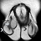
A rare case
of extratesticular scrotal epidermoid cyst. MR axial T1 weighted image showed an isointense lesion (blue arrow) adjacent to the right epididymal head with a central hypointense focus.

A rare case
of extratesticular scrotal epidermoid cyst. MR axial T2 weighted image showed a hypointense lesion (blue arrow) adjacent to the right epididymal head with a central hypointense focus.

A rare case
of extratesticular scrotal epidermoid cyst. MR coronal T2- STIR image showed a hypointense lesion adjacent to the right epididymal head with a central hypointense focus.

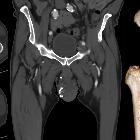
Phlebolithen
in skrotaler Varicocele: Computertomografie, 10 mm MIP koronar.


 Assoziationen und Differentialdiagnosen zu skrotale Verkalkungen: