extratestikuläre intraskrotale Epidermoidzyste

Extratesticular
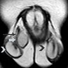
epidermal inclusion cyst of the scrotum (ECR 2016 Case of the Day). Coronal T2-weighted image shows right extratesticular mass (arrow), close to the ipsilateral spermatic cord (long arrow), mainly hyperintense, with signal similar to that of normal testes (arrowheads), surrounded by a low signal intensity capsule.

Extratesticular
epidermal inclusion cyst of the scrotum (ECR 2016 Case of the Day). Transverse T1-weighted image depicts homogeneous right extratesticular mass (arrow), isointense to the ispilateral testis (arrowhead).

Extratesticular
epidermal inclusion cyst of the scrotum (ECR 2016 Case of the Day). Transverse sonographic image of the right hemiscrotum depicts a solid, heterogeneous extratesticular mass (arrow). The lesion is sharply defined, measuring 23 mm in maximal diameter.

Extratesticular
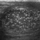
epidermal inclusion cyst of the scrotum (ECR 2016 Case of the Day). Sagittal sonographic image of the scrotum shows right extratesticular mass, clearly separated from the ipsilateral testis (long arrow). The lesion is hypoechoic, with multiple internal echogenic foci (arrowheads).

Extratesticular
epidermal inclusion cyst of the scrotum (ECR 2016 Case of the Day). Colour Doppler sonographic image depicts no internal Doppler signals (arrow).

Extratesticular
epidermal inclusion cyst of the scrotum (ECR 2016 Case of the Day). Transverse ADC (b= 900 s/mm2) shows lesion’s restricted diffusion (long arrow), due to the presence of keratin debris histologically. The ADC of the mass is 0.48 × 10⁻³mm2/s [right testis (arrowhead), 1.02 × 10⁻³mm2/s].

Extratesticular
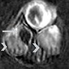
epidermal inclusion cyst of the scrotum (ECR 2016 Case of the Day). Coronal dynamic contrast-enhanced subtracted image at early phase (120 seconds) depicts absence of lesion enhancement (long arrow), a finding strongly suggestive of benignity. Both testes (arrowheads) enhance moderately and homogeneously.

A rare case
of extratesticular scrotal epidermoid cyst. A non-vascular lesion measuring 15x12 mm was noted indenting the lateral aspect of the right epididymal head with a whorled appearance and central calcific focus giving it a "target" or "bulls eye" appearance.

A rare case
of extratesticular scrotal epidermoid cyst. The lesion showing a whorled appearance with a central echogenic focus suggestive of calcification.

A rare case
of extratesticular scrotal epidermoid cyst. MR axial T1 weighted image showed an isointense lesion (blue arrow) adjacent to the right epididymal head with a central hypointense focus.

A rare case
of extratesticular scrotal epidermoid cyst. MR axial T2 weighted image showed a hypointense lesion (blue arrow) adjacent to the right epididymal head with a central hypointense focus.

A rare case
of extratesticular scrotal epidermoid cyst. MR coronal T2- STIR image showed a hypointense lesion adjacent to the right epididymal head with a central hypointense focus.
extratestikuläre intraskrotale Epidermoidzyste
Siehe auch:
und weiter:

 Assoziationen und Differentialdiagnosen zu extratestikuläre intraskrotale Epidermoidzyste:

