Tumoren des Skrotums
Extratesticular
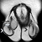
epidermal inclusion cyst of the scrotum (ECR 2016 Case of the Day). Coronal T2-weighted image shows right extratesticular mass (arrow), close to the ipsilateral spermatic cord (long arrow), mainly hyperintense, with signal similar to that of normal testes (arrowheads), surrounded by a low signal intensity capsule.

Extratesticular
epidermal inclusion cyst of the scrotum (ECR 2016 Case of the Day). Transverse T1-weighted image depicts homogeneous right extratesticular mass (arrow), isointense to the ispilateral testis (arrowhead).

Leiomyosarcoma
of the spermatic cord: a rare paratesticular neoplasm case report. USS left groin demonstrating a well-circumscribed ovoid, solid, and vascular lesion, with heterogeneous internal echotexture

Left
paratesticular rhabdomyosarcoma in 15-year-old male.. Gray scale ultrasound of the left hemiscrotum in the region of the epididymal body reveals a large, markedly heterogeneous, non calcified mass involving the epididymal body.


 Assoziationen und Differentialdiagnosen zu Tumoren des Skrotums:

