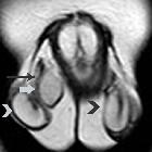
Rhabdomyosarkom des Skrotums


Rhabdomyosarkom des Skrotums
Rhabdomyosarkom des Urogenitaltraktes Radiopaedia • CC-by-nc-sa 3.0 • de
Rhabdomyosarcomas of the genitourinary tract are uncommon tumors occurring in pelvic organs. It is a disease nearly exclusive to the pediatric population.
For a general discussion of this type of tumor, please refer to the article on rhabdomyosarcomas.
Epidemiology
The peak incidence of tumors is between 3-6 years of age, with a slight male predominance of 2-3:1 .
Prostatic origin is the most common in males. The bladder is the most frequent site which affects both sexes.
The vagina, cervix and uterus may all be involved in females. The vagina is the most prevalent in the female genital tract .
Paratesticular tumors are the only genitourinary tract rhabdomyosarcomas that tend to occur in older children, typically adolescents.
Clinical presentation
A pelvic or scrotal mass is the most common complaint, with or without pain. The mass is typically large at presentation. Urinary symptoms may be a feature, such as hematuria and dysuria .
The symptoms depend, to an extent, on which organ is the site for the tumor.
Pathology
As with rhabdomyosarcomas at other sites, there are 4 major subtypes; embryonal, botryoid variant of embryonal type, alveolar and undifferentiated.
The embryonal cell type is most common, accounting for more than half of all histological subtypes.
The botryoid variant is the most frequent to affect the bladder.
Radiographic features
Ultrasonography
Usually the first imaging investigation given its ubiquitous use in the pediatric population. A heterogeneous mass, with variable solid and cystic elements, is often encountered. Doppler interrogation may demonstrate internal vascular flow.
CT
A heterogeneous mass as with ultrasound. Its relationship to other pelvic organs is delineated, in particular, to review for local invasion.
Used to assess for locoregional lymphadenopathy and distant metastatic disease in the lungs, liver and bones.
MRI
In the pelvis may add further to CT in delineated the tumor's relationship to adjacent organs and identifying lymph node disease.
Treatment
Primary surgery with both chemotherapy and radiotherapy play a role in management.
Siehe auch:

 Assoziationen und Differentialdiagnosen zu Rhabdomyosarkom des Skrotums:


