Rhabdomyosarkom


Rhabdomyosarcoma (RMS) is a malignant tumor with skeletal muscle cell morphology. It is one of the tumors of muscular origin.
This article focuses on a general discussion of rhabdomyosarcomas. For location specific details, please refer to:
- rhabdomyosarcomas of the biliary tract
- rhabdomyosarcomas of the genitourinary tract
- rhabdomyosarcoma of the heart
- rhabdomyosarcomas of the head and neck
- rhabdomyosarcomas of the orbit
Epidemiology
Rhabdomyosarcomas are the most common soft tissue tumor in children and account for 5-8% of childhood cancers, and 19% of all pediatric soft tissue sarcomas .
In general, they are found in young patients, less than 45 years of age , with ~65% diagnosed in patients under 10 years old .
There is a slight male predilection (M:F 1.67:1 ) with Caucasian children affected more often than children of other races.
Clinical presentation
Clearly, clinical presentation will vary depending on the location of a tumor (see below); however, in general, rhabdomyosarcomas are rapidly growing masses. They cause localized pressure effects on neurovascular structures and have a predilection for infiltrating bones . Pathological fractures could therefore occur.
These tumors can occur anywhere, and not necessarily where the skeletal muscle is normally found. In children and adolescents, they occur predominantly in the head, neck and pelvis.
Distribution
Rhabdomyosarcomas are found essentially anywhere in the body :
- head and neck: ~50% *
- orbit: ~20%
- oropharynx/nasopharynx, palate: ~15%
- sinuses, mastoid, middle ear: ~15%
- genito-urinary: ~25%
- paratesticular: ~20%
- bladder: ~5%
- extremities: ~15%
- other: ~10%
- trunk and thorax: 7%
- gastrointestinal tract: 1%
*: see note on figures/percentages
Pathology
Rhabdomyosarcomas are thought not to arise from skeletal muscle, but rather to differentiate into a tumor which resembles skeletal muscle . This accounts for it arising in locations where no skeletal muscle is present. It is divided into three subtypes :
- spindle cell rhabdomyosarcoma: 50-66%
- botryoid rhabdomyosarcoma: 5-10% (best prognosis)
- anaplastic rhabdomyosarcoma
Associations
Although the vast majority of cases are sporadic, increased incidence of rhabdomyosarcomas is seen in patients with a variety of syndromes and congenital anomalies, including :
- neurofibromatosis type I (NF1)
- Beckwith-Wiedemann syndrome
- Li-Fraumeni syndrome
- DICER1 syndrome
- Costello syndrome
- maternal use of cocaine and marijuana
Staging
Please refer to Rhabdomyosarcoma staging.
Radiographic features
Unfortunately, the appearance of the mass itself is non-specific and indistinguishable from other sarcomas. The location and demographics of the patient are most useful in narrowing the differential.
Plain radiograph
Although entirely non-specific plain films are a useful first step as they can give a quick global view of the region and identify calcifications in the mass, bony involvement and metastases. The mass appears of soft tissue density.
When present in the extremities in children, embryonal rhabdomyosarcomas may cause bowing of the adjacent long bones. This should not be thought of as suggesting slow growth or indolent behavior .
Ultrasound
- heterogeneous well-defined irregular mass of low to medium echogenicity
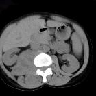
CT
- soft tissue density
- some enhancement with contrast
- adjacent bone destruction is seen in over 20% of cases
MRI
Signal characteristics include:
- T1
- low to intermediate intensity, isointense to adjacent muscle
- areas of hemorrhage are common in alveolar and pleomorphic subtypes
- T2
- hyperintense
- prominent flow voids may be seen particularly in extremity lesions
- T1 C+ (Gd): shows considerable enhancement
Embryonal rhabdomyosarcomas tend to be more homogeneous, whereas alveolar and pleomorphic rhabdomyosarcomas frequently have areas of necrosis . The latter is associated with ring-like enhancement .
Treatment and prognosis
Unfortunately, up to 20% of patients have metastases at the time of diagnosis . These are typical to lung and bone marrow.
Treated with combination surgery, chemotherapy, and radiation:
- surgery: resection of a primary tumor, if necessary after down-staging chemoradiotherapy
- chemotherapy: common agents include vincristine, cyclophosphamide, dactinomycin, adriamycin, ifosfamide, VP-16
- radiotherapy: external beam radiation is used in some cases of rhabdomyosarcoma
Survival varies dependent on primary location, histological type, local invasion and metastases. Overall 5-year survival is approximately 75% .
Siehe auch:
- Neurofibromatose Typ 1
- Beckwith-Wiedemann-Syndrom
- botryoid rhabdomyosarcoma
- Rhabdomyosarkom der Kopf-Hals-Region
- pleomorphic rhabdomyosarcoma
- alveolar rhabdomyosarcoma
- embryonales Rhabdomyosarkom
- tumours of muscular origin
- tumours of the masticator space
- rhabdomyosarcoma staging
- Rhabdomyosarkom der Blase
- retroperitoneales Rhabdomyosarkom
- rhabdomyosarcomas of the biliary tract
- Spindelzelliges Rhabdomyosarkom
- anaplastic rhabdomyosarcoma
- note on figures / percentages
und weiter:
- kongenitale pulmonale Atemwegsmalformation (CPAM)
- Neuroblastom
- WHO-Klassifikation der Tumoren des zentralen Nervensystems
- Ästhesioneuroblastom
- Steißbeinteratom
- Hepatoblastom
- radiologisches muskuloskelettales Curriculum
- Knochentumoren
- Proteus-Syndrom
- Hydrokolpos
- Tumoren der Thoraxwand
- kapilläres Hämangiom der Orbita
- Weichteilsarkom
- Rhabdomyosarkom der Orbita
- Metastasen im Hoden
- Hydrometrokolpos
- rhabdomyosarcoma of the orbit
- small round blue cell tumours
- Vergrößerung der Glandula parotis
- malignancies in childhood
- muscular tumours
- Sarcoma botryoides
- malignant liver tumours (paediatric)
- fetale Tumoren
- maligner rhabdoider Tumor
- primary malignancies of the nasopharynx
- metastatic nasopharyngeal rhabdomyosarcoma
- Teratom des Pharynx
- paediatric bone tumours (differential diagnosis)
- primäre nasopharyngeale maligne Tumoren
- alveoläres Rhabdomyosarkom (Unterschenkel)
- entdifferenziertes Chondrosarkom
- Plattenepithelkarzinom der Mundhöhle
- Rhabdomyosarkom des Urogenitaltraktes
- Rhabdomyosarkom der Prostata
- Rhabdomyosarkom des Skrotums
- paratestikuläre Tumoren
- metastatic rhabdomyosarcoma
- rhabdomyosarcoma of the psoas muscle
- primäres Lymphom der Muskulatur


 Assoziationen und Differentialdiagnosen zu Rhabdomyosarkom:

