Gilula three carpal arcs

Piece of pie
sign (wrist) • Gilula carpal arcs - Ganzer Fall bei Radiopaedia

Gilula three
carpal arcs • Gilula carpal arcs (diagram) - Ganzer Fall bei Radiopaedia
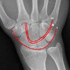
Gilula three carpal arcs refer to the carpal alignment described on posteroanterior or anteroposterior wrist radiographs and are used to assess normal alignment of the carpus:
- first arc: is a smooth curve outlining the proximal convexities of the scaphoid, lunate and triquetrum
- second arc: traces the distal concave surfaces of the same bones
- third arc: follows the main proximal curvatures of the capitate and hamate
Alignment
- carpal bones have smooth and rounded edges to varying degrees, lines joining these convexities form arcs, when major convexities are used in drawing
- there should be no step-offs in the contour of the arcs, except for two normal variants
- a triquetrum that is shorter than the lunate creates a step-off in the first arc but there is still a normal second arc
- "bi-lobed" appearance of second carpal arc in lunate type II morphology
- disrupted arc may indicate a ligamentous injury or fracture at the site of the broken arc
History and etymology
The concept of three radiographic arcs was first proposed by Louis A Gilula (1942-2014) in 1979 .
Siehe auch:
und weiter:


 Assoziationen und Differentialdiagnosen zu Gilula-Linien:
