Scapholunäre Dissoziation


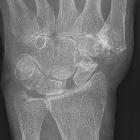
Scapholunate dissociation, also known as rotary subluxation of the scaphoid, refers to an abnormal orientation of the scaphoid relative to the lunate, and implies severe injury to the scapholunate interosseous ligament and other stabilizing ligaments.
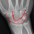
Carpal dissociation implies carpal instability, which has important clinical implications; thus it is essential to identify this finding on imaging. Note that absence of dissociation does not exclude ligamentous injury, as lower grade injuries which result in dynamic instability may present with normal radiographic carpal alignment.
The typical pattern of scapholunate dissociation consists of:
- relative flexion (volar rotation) of the scaphoid
- relative extension (dorsal rotation) of the lunate
Terminology
Although "scapholunate dissociation" is a descriptive term, it is used synonymously with its primary pathophysiologic correlate, scapholunate interosseous ligament (SLIL) injury. Although injury to additional ligaments is present in scapholunate dissociation, the SLIL is the major target of surgical reconstruction.
Dorsal intercalated segment instability (DISI) is a related carpal malalignment pattern which also results from rupture of the scapholunate interosseous ligament .
Epidemiology
Scapholunate dissociation most commonly results from trauma. It is the leading cause of SLAC (scapholunate advanced collapse) wrist, which is the most common pattern of degenerative arthritis in the wrist .
Clinical presentation
Scapholunate dissociation usually presents following a fall with minimal swelling and pain localized over the dorsal scapholunate region. Presentation is often delayed in the absence of an associated fracture. Pain is increased by dorsiflexion.
Pathology
The scapholunate interosseous ligament (SLIL) is a U-shaped ligament which is arbitrarily divided into three anatomic components: dorsal, intermediate and volar :
- dorsal component:
- strongest, most important in resisting volar-dorsal translation
- 3 mm in thickness and composed of short, transversely-oriented collagen fibers
- intermediate component:
- primarily composed of fibrocartilage, homologous to the meniscus of the knee
- volar component:
- 1 mm in thickness
Major injury of the SLIL (complete tear of dorsal component) and radiolunate ligament may result in scapholunate dissociation. However, even complete SLIL transection does not result in permanent dissociation due to the presence of the secondary scaphoid stabilizers, e.g. palmar radioscaphoid-capitate, scaphoid capitate, and anterolateral scapho-trapezio-trapezoid ligaments. Long-term wear on these secondary ligamentous stabilizers may lead to permanent deformity / scapholunate advanced collapse .
Mayfield et al. have proposed a four-stage process to describe perilunar wrist instability, in which scapholunate dissociation represents stage 1 .
Radiographic features
Plain radiograph
- widened scapholunate interval > 4 mm on PA view (i.e. Terry Thomas sign)
- scapholunate interval should be measured at the midpoint of the adjacent parallel articular contours of the two bones (the scapholunate interval normally narrows proximal to distal)
- widening is exacerbated on
- clenched fist views
- PA views with wrist in ulnar deviation (causes intercalated segment extension)
- increased scapholunate angle on lateral view
- scaphoid rotates into flexion, which will often increase the scapholunate angle to greater than 60 degrees
- exaggerated cortical ring distal scaphoid on AP view (i.e. signet ring sign)
- ringed appearance of distal scaphoid, resulting from the distal pole viewed en face
- shortening of interval between the cortical ring and the proximal margin of scaphoid < 7 mm
Treatment and prognosis
Acute non-displaced and chronic asymptomatic SLIL injuries may be treated conservatively with non-steroidal anti-inflammatories (NSAIDs) and immobilization .
Surgical repair or reconstruction of the scapholunate interosseous ligament is normally required to prevent long-term complications, namely proximal migration of the capitate between the scaphoid and lunate with a resultant degenerative disease known as SLAC wrist (scapholunate advanced collapse).
Differential diagnosis
- dorsal intercalated segmental instability
- scapholunate angle to greater than 60 degrees and capitolunate angle greater than 30 degrees
- volar intercalated segmental instability
- scapholunate angle to lesser than 30 degrees and capitolunate angle greater than 30 degrees
- scapholunate advanced collapse
- a consequence of undiagnosed or untreated scapholunate ligament injury resulting in radioscaphoid malalignment, progressive chondromalacia, and osteoarthritis
Siehe auch:
- DISI-Fehlstellung
- Gilula-Linien
- Terry-Thomas sign
- perilunäre Luxation
- scapholunate advanced collapse (SLAC)
- karpale Luxationen
- Mayfield-Klassifikation
- Ligamentum scapholunatum
und weiter:

 Assoziationen und Differentialdiagnosen zu Scapholunäre Dissoziation:


