scapholunate advanced collapse (SLAC)


Scapholunate advanced collapse (SLAC), commonly known as SLAC wrist, refers to a pattern of wrist malalignment that has been attributed to post-traumatic or spontaneous osteoarthritis of the wrist. It is a complication that can occur with undiagnosed or untreated scapholunate dissociation. It is essentially the same sequela of wrist injury causing scaphoid nonunion as seen in scaphoid nonunion advanced collapse (SNAC).
Pathology
SLAC is most commonly a consequence of undiagnosed or untreated scapholunate ligament injury and rotatory subluxation of the scaphoid bone resulting in radioscaphoid malalignment, progressive chondromalacia, and osteoarthritis.
Its features, however, also have been observed in patients with idiopathic calcium pyrophosphate dihydrate (CPPD) crystal deposition disease.
Radiographic features
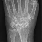
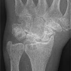
The pattern is that of progressive osteoarthritis affecting initially the articulation between the radial styloid and the scaphoid. In later stages of the disease, osteoarthritis affects the whole radioscaphoid articulation, then the articulation between lunate and capitate. Finally, it may involve other intercarpal joints. In addition, there is scapholunate interval widening as well as proximal migration of the scaphoid and the capitate .
Classification
Watson staging is often used by hand surgeons :
- I: osteoarthritis of the articulation between the radial styloid and the scaphoid
- II: osteoarthritis involving the whole radioscaphoid articulation
- III: osteoarthritis of the radioscaphoid and capitolunate articulations
- IV: osteoarthritis of the radiocarpal and intercarpal articulations +/- distal radioulnar joint (DRUJ)
Note that the radiolunate joint is almost preserved until the very last stages of the disease. It is also worth noting that the scaphoid fossa in the radius may be deep/preserved in cases of CPPD in contrast to post-traumatic SLAC wrist.
Plain radiograph
Key assessing parameters include
- scapholunate diastasis (PA radiograph)
- rotatory subluxation of the scaphoid (lateral radiograph)
- features associated with dorsal intercalated segment instability (DISI) (lateral radiograph)
CT
Sagittal reformatted images from multidetector CT arthrographic data are particularly useful in demonstrating abnormal angulations of the scaphoid and lunate bones (increased scapholunate angle and dorsal or volar intercalated segment instability deformity), radioscaphoid incongruity, cartilage loss, and subchondral bone degenerative changes.
Treatment and prognosis
Surgical treatment for SLAC wrist includes four-corner arthrodesis, capitolunate arthrodesis, complete wrist arthrodesis, scaphoidectomy, proximal row carpectomy (PRC), denervation, and radial styloidectomy .
Siehe auch:
- Scapholunäre Dissoziation
- scaphoid non-union advanced collapse
- Kalziumpyrophosphat-Ablagerungskrankheit
- posttraumatische Kahnbeinnekrose
- Knocheninfarkt Scaphoid
und weiter:


 Assoziationen und Differentialdiagnosen zu scapholunate advanced collapse (SLAC):