Arthrose Handwurzel

Fortgeschrittene
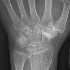
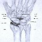
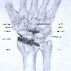
Arthrose zwischen dem Scaphoid einerseits und dem Trapezium und Trapezoideum andererseits (STT-Arthrose) mit aufgehobenem Gelenkspalt, erheblicher Sklerosierung und periartikulären Verkalkungen. Oft mit einer Rhizarthrose vergesellschaftet, die in diesem Fall nur deutlich geringer ausgeprägt ist.

Severe
osteoarthritis and osteopenia of the carpal joint and 1st carpometacarpal joint.


Scapholunate
advanced collapse: a pictorial review. Coronal illustrations of the wrist depicting the progressive osteoarthritic changes of SLAC arthropathy. a Normal wrist demonstrating an intact scapholunate ligament (white arrowhead), normal scapholunate interval and preservation of the joint spaces. (S scaphoid, L lunate, C capitate, R radius, U ulna). b Stage I: Early findings of SLAC arthropathy including scapholunate ligament tear (black arrowhead) with mild widening of the scapholunate interval, as well as early osteoarthritic changes involving the most radial portion of the radioscaphoid joint (black arrow). c Stage II: Progression of SLAC arthropathy with worsening osteoarthritic changes which now involve the entire radioscaphoid articulation (black arrows), and there has been increased widening of the scapholunate interval. d Stage III: Further progression of SLAC arthropathy depicted by worsening radioscaphoid joint osteoarthritic changes (black arrows), and there is now narrowing of the capitolunate joint space with associated osteoarthritic changes (white arrow). This will eventually progress to further proximal migration of the capitate

Scapholunate
advanced collapse: a pictorial review. SLAC wrist early radiological findings (Stage I). Posteroanterior radiograph of the wrist (a), coronal CT reformat image of the wrist (b) and coronal proton density fat suppressed MRI image of the wrist (c) in three different patients show spurring at the articular/non-articular junction on the radial side of the scaphoid (black arrows) and at the radial styloid tip (white arrows), with loss of the normal rounded curvature of the radial styloid. Scapholunate diastasis is seen (black asterisks)

Scapholunate
advanced collapse: a pictorial review. SLAC wrist radiological progression (Stage II). Posteroanterior radiograph (a), coronal CT reformat image (b) and coronal T1-weighted MRI image of the wrists (c) of three different patients, demonstrating narrowing of the radioscaphoid articulation (black arrows) in addition to radial styloid and scaphoid spurring. Note the preservation of the capitolunate joint at this point (white arrows)

Scapholunate
advanced collapse: a pictorial review. SLAC wrist with further radiological progression (Stage III). Posteroanterior radiograph (a) and coronal and sagittal CT reformat images (b) of the same patient, as well as a coronal STIR MRI image of the wrist (c) in a different patient with SLAC wrist arthropathy. Images demonstrate narrowing of the capitolunate joint (black arrows) in addition to involvement of the radioscaphoid joint. Note preservation of the radiolunate joint (white arrows). There is no significant migration of the capitate at this stage
 Assoziationen und Differentialdiagnosen zu Arthrose Handwurzel:


