Xanthom
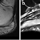
Imaging of
plantar fascia disorders: findings on plain radiography, ultrasound and magnetic resonance imaging. Plantar xanthoma. On both sagittal T1-weighted (a) and fluid-sensitive (b) images, xanthoma (arrows) appears as fusiform enlargement of the PF and shows heterogeneous signal intensity

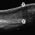
Multiple
tendon xanthomas in a child with familial hypercholesterolaemia. Panoramic ultrasound image of right achilles tendon along its long axis (arrows). Diffuse thickening of achilles tendon is noted with loss of fibrillary architecture.
Multiple
tendon xanthomas in a child with familial hypercholesterolaemia. Panoramic ultrasound image of left Achilles tendon along its long axis (arrows). Diffuse thickening of achilles tendon is noted with loss of fibrillary architecture.
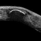
Multiple
tendon xanthomas in a child with familial hypercholesterolaemia. Panoramic ultrasound image of right knee sagittal section. Quadriceps tendon (hollow arrows) and patellar tendon (solid arrows) show fusiform thickening with loss of fibrillary architecture.
Multiple
tendon xanthomas in a child with familial hypercholesterolaemia. Panoramic ultrasound image of left knee sagittal section. Quadriceps tendon (hollow arrows) and patellar tendon (solid arrows) show fusiform thickening with loss of fibrillary architecture.
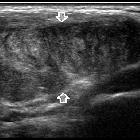
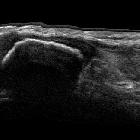
Multiple
tendon xanthomas in a child with familial hypercholesterolaemia. High resolution ultrasound of right patellar tendon in short axis (arrows). Thickening of tendon is noted with convex inner margin and altered nodular echotexture.


 Assoziationen und Differentialdiagnosen zu Xanthom: