venous tumors


Primary
leiomyosarcoma of inferior vena cava. Lobulated mass within the suprarenal and intrahepatic segments of IVC, distending the vein. The mass stops short of the hepatic veins.

Primary
leiomyosarcoma of inferior vena cava. Hypervascular mass anterior and to the right of abdominal aorta. The lesion is inseparable from the IVC. Note the dilated subcutaneous veins in anterior abdominal wall.

Primary
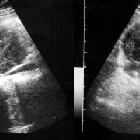
leiomyosarcoma of inferior vena cava. Hypoechoeic mass seen medial to right renal hilum involving renal vein and IVC.

Primary
leiomyosarcoma of inferior vena cava. Large lobulated heterogeneously enhancing mass involving lower and mid segments of IVC extending into right renal vein. The mass stops short of the hepatic veins. Thrombus is also seen in distal IVC.

Primary
leiomyosarcoma of inferior vena cava. Heterogeneously enhancing retroperitoneal mass which is in the location of IVC. Note the dilated collateral veins posterior to liver.

Primary
leiomyosarcoma of inferior vena cava. Lobulated, enhancing mass in the IVC with extraluminal component anteriorly. A bland thrombus is also seen in distal IVC.

Symplastic
Leiomyoma in the Suprarenal Inferior Vena Cava:A. Coronal and B. Axial MRI following IV injection of contrast material shows a moderate level of heterogeneous contrast enhancement when compared to the surrounding muscle planes. The mass could not be separated from the inferior vena cava.
 Assoziationen und Differentialdiagnosen zu Tumoren der Venen:


