Leiomyom der Vena cava

Symplastic
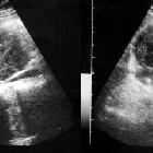
Leiomyoma in the Suprarenal Inferior Vena Cava:US imaging, heterogeneous echogenicity and lobulated contour mass

Symplastic
Leiomyoma in the Suprarenal Inferior Vena Cava:Axial T1-weighted MR image shows isointensity of the mass compared to the skeletal muscle.

Symplastic
Leiomyoma in the Suprarenal Inferior Vena Cava:A. Coronal and B. Axial MRI following IV injection of contrast material shows a moderate level of heterogeneous contrast enhancement when compared to the surrounding muscle planes. The mass could not be separated from the inferior vena cava.
Leiomyom der Vena cava
Leiomyom Radiopaedia • CC-by-nc-sa 3.0 • de
Leiomyoma is a benign smooth muscle (myometrial) tumor, most commonly found in the uterus.
Classification
Leiomyoma is classified by location:
- uterine leiomyoma
- cervical leiomyoma
- leiomyoma of the urinary bladder
- urethral leiomyoma
- solitary cutaneous leiomyoma
- vascular leiomyoma (angioleiomyoma)
- dartoic leiomyoma - arises from the dartos muscle of the nipple or genitals.
- esophageal leiomyoma
- leiomyoma of the jejunum
See also
- leiomyosarcoma
- angiolipoleiomyoma
- diffuse uterine leiomyomatosis
- diffuse peritoneal leiomyomatosis
- esophageal leiomyomatosis
- hereditary leiomyomatosis and renal cell carcinoma
- intravenous leiomyomatosis
- benign metastasizing leiomyoma
- parasitic leiomyoma
- extra-uterine pelvic leiomyoma
Siehe auch:

 Assoziationen und Differentialdiagnosen zu Leiomyom der Vena cava:

