epithelioid sarcoma

Young adult
treated for leukemia as a preschooler, now with new palpable sternal massAxial CT without contrast of the chest with soft tissue (above) and lung (below) windows shows a heterogeneous mass contiguous with the rectus muscles and anterior to the lower sternum which appears to be eroding into the sternum along with multiple peripheral pulmonary soft tissue nodules.The diagnosis was epithelioid sarcoma with pulmonary metastases.

Epithelioid
sarcoma in the chest wall: a case report and literature review. Enhanced chest CT shows a right chest wall tumor with calcification (white arrow) (a). Chest MRI shows an invasive dumbbell-shaped tumor, which is isointense on T1-weighted images (b) and heterogeneously hyperintense on T2-weighted images (c)

Achilles
tendon reconstruction with peroneus tendon transfer following epithelioid sarcoma resection: a rare case report at 5 years follow-up. Pre-operative MRI showed a mass in the context of the middle third of Achilles tendon, with hyperintense signal in T2-weighted and T2-weighted fat-sat images

Primary
pulmonary epithelioid sarcoma: a case report. Chest computed tomographic scan showing a 2.0-cm soft-tissue nodule in the right upper lobe of the lung

Epithelioid
sarcoma with muscle metastasis detected by positron emission tomography. Epithlioid sarcoma in the forearm. Flexion contracture of the fingers can be seen (A). Plain radiograph shows irregular surface of the ulna (B). MRI of the forearm shows an abnormal lesion with iso-signal intensity on T1-weighted image (top) and high-signal intensity on T2-weighted image (middle) (C). Enhancement with gadolinium can be seen on T1-weighted fat-suppression image (bottom) (C).

Epithelioid
sarcoma with muscle metastasis detected by positron emission tomography. Metastatic lesions of epithelioid sarcoma. FDG-PET identifies lesions with an increased uptake in the right neck (SUVmax; 4.6), right upper arm (SUVmax; 4.1 g/ml), left upper arm (SUVmax; 4.2 g/ml), right thigh (SUVmax; 5.5 g/ml) left thigh (SUVmax; 2.0 g/ml), back (SUVmax; 3.6 g/ml), and lower back (SUVmax; 4.6 g/ml), as well as the left axilla (SUVmax; 3.9 g/ml) (arrows) (A). CT with contrast medium shows a swollen axillary lymph node (B). Sagittal MRI section of the right thigh shows a nodular lesion associated with prominent longitudinal abnormal signal intensity on T2-weighted image (C). The nodular lesion shows iso-intensity on T1-weighted fat-suppression image (top), and heterogeneous high-intensity on T2-weighted image (middle) (D). Enhancement with gadolinium can be seen on T1-weighted fat-suppression image (bottom) (D).

Delayed
diagnosis of pediatric intra-articular epithelioid sarcoma: a case report and literature review. The MRI images of the right knee from different positions. (a1-d1) In the first hospitalization, the MRI images showed a large amount of fluid signal shadow in the right knee joint cavity from different positions, including the lateral sagittal position (a1, T1WI; c1 PDWI), the transverse axial position (b1, T2WI) and the coronary position (d1, PDWI). (a2-d2) During the second hospitalization, the MRI images highlighted patchy high signal shadows (white arrows) in the surrounding soft tissues of the right knee (observed in a2, T1WI; b1, T2WI; c2 PDWI; d2 PDWI-COR). A patchy low T1-weighted signal (a2) and high PD-weighted fat-suppressed signal (c2, d2) were seen in the distal metaphysis of the right femur and proximal metaphysis of the tibia, with unclear borders. Fluid signal shadows and strip-like signal shadows were seen within the right knee joint cavity. Abbreviations: T1WI, T-weighted image; T2WI, T2-weighted image; PDWI, proton density-weighted image; PDWI-COR, coronary proton density-weighted image

Epithelioid
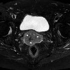
sarcoma of the uterine cervix. Axial (b & fat-suppressed c) T2-weighted images confirmed predominantly exophytic lower cervical mass (*) with moderate-to-high signal intensity, 52x36mm axial diameters. Note absent pelvic lymphadenopathies and effusion in peritoneal cul-de-sac.
Das Epitheloidsarkom ist ein seltenes, aber aggressives Weichteilsarkom mit epitheloider Zellmorphologie und häufig ungünstiger Prognose aufgrund von Lokalrezidiven und Metastasen. Häufige Lokalisationen sind die distalen Extremitäten, jedoch sind auch andere Fälle zum Beispiel an den Organen des Beckens beschrieben.
Siehe auch:
und weiter:

 Assoziationen und Differentialdiagnosen zu Epitheloidsarkom:

