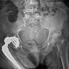
Komplikationen bei Hüftendoprothesen

Luxiertes
Inlay einer Hüftendoprothese: Der Prothesenkopf steht dezentriert im Rest der Pfanne, das Inlay liegt dorsal davon im Weichteil (in der axialen Schicht als Bogen sichtbar).

Hüft-Spacer
(Synicem) als temporärer Platzhalter bei infizierter Hüftendoprothese. Wenn die Infektion ausgeheilt ist, wird wieder eine dauerhafte Hüftendoprothese implantiert.


Luxiertes
Inlay einer Hüftendoprothese: Der Prothesenkopf steht dezentriert im Rest der Pfanne, das Inlay liegt dorsal davon im Weichteil (in der axialen Schicht als Bogen sichtbar).

Complications
of total hip arthroplasty • Hip prosthesis - Ganzer Fall bei Radiopaedia

Complications
of total hip arthroplasty • Loose right total hip prosthesis - Ganzer Fall bei Radiopaedia

Complications
of total hip arthroplasty • Hip prosthesis dislocation - Ganzer Fall bei Radiopaedia

Complications
of total hip arthroplasty • Hip prosthesis dislocation - Ganzer Fall bei Radiopaedia

Complications
of total hip arthroplasty • Cortical hypertrophy in total hip arthroplasty with cementless stem - Ganzer Fall bei Radiopaedia

Complications
of total hip arthroplasty • Fractured femoral prosthetic stem - Ganzer Fall bei Radiopaedia

Complications
of total hip arthroplasty • Dislocated tumor prosthesis - Ganzer Fall bei Radiopaedia

Complications
of total hip arthroplasty • Periprosthetic fracture - Ganzer Fall bei Radiopaedia

Complications
of total hip arthroplasty • Dislocated right hip prosthesis - Ganzer Fall bei Radiopaedia

Complications
of total hip arthroplasty • Dislocation of hip replacement - Ganzer Fall bei Radiopaedia

Complications
of total hip arthroplasty • Acetabular protrusion - Ganzer Fall bei Radiopaedia

Metal
artifact reduction sequence • Abductor tendon tear and bursitis after total hip arthroplasty - Ganzer Fall bei Radiopaedia

Röntgenbild
eines rechten Hüftgelenks mit zementfreier Endoprothese (Rechteckschaft und konische Schraubpfanne): Konzentrische Einstellung des Metallkopfs im Polyethylen-Inlay der Pfanne. Ausgeprägte heterotope periartikuläre Verknöcherung und massive Osteolyse in der Umgebung des Prothesenschafts. Bei operativer Revision massive Metallose.

Complications
of total hip arthroplasty • Loosening of hip prosthesis - Ganzer Fall bei Radiopaedia


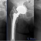
Heterotope
periprothetische Ossifikation: links postoperativ, Mitte und rechts nach 5 Monaten.

Girdlestone-Situation
nach Ausbau einer infizierten Totalendoprothese des linken Hüftgelenks. Zur Zemententfernung wurde das obere Femurende gefenstert. Der Kortikalisspan ist mit drei Cerclagen refixiert. Im Pfannenlager und in der Markhöhle des Femurs sind PMMA-Ketten eingelegt.

Hüftendoprothese
mit defektem Inlay, zumindest disloziert, mutmaßlich gebrochen. Der Prothesenkopf ist deutlich nach kranial lateral dezentriert.

Bruch des
Prothesenkopfes bei Hüft-Totalendoprothese: Linkes Bild mit intakter Prothese, aber schon reichlich periarthikuläre Ossifikationen. Rechtes Bild Fragmentierung des Prothesenkopfes. Die Ossifikationen haben auch zugenommen.

Hip
dislocation • Dislocated hip prosthesis - Ganzer Fall bei Radiopaedia

Acetabular
protrusion • Protrusio acetabuli - Ganzer Fall bei Radiopaedia

Particle
disease • Particle disease - total hip replacement - Ganzer Fall bei Radiopaedia

Complications
of total hip arthroplasty • Dislocated total hip replacement - Ganzer Fall bei Radiopaedia

Complications
of total hip arthroplasty • Hip prosthesis loosening - Ganzer Fall bei Radiopaedia
Komplikationen bei Hüftendoprothesen
Siehe auch:
- Protrusio acetabuli
- Pseudobursa
- periprothetische Ossifikationen Hüftgelenk
- Metallose
- Hüftendoprothese
- Prothesenlockerung
- Luxation Hüftprothese
- Prothesenlockerung Hüftendoprothese
- Protheseninfektion
- hip replacement stress shielding
- infizierte Hüftprothesen
- aggressive zystische Granulomatose bei Hüftprothese
- Komplikationen bei Knieendoprothesen
- Materialkomplikationen bei Hüftendoprothesen
und weiter:

 Assoziationen und Differentialdiagnosen zu Komplikationen bei Hüftendoprothesen:
aggressive
granulomatosis post hip replacement


