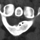
Kompressionssyndrom Nervus ulnaris

The elbow:
review of anatomy and common collateral ligament complex pathology using MRI. A 60-year-old woman with pain, muscle weakness, and paresthesias. Axial FS PD-weighted MRI (a, b) showing an enlargement and hyperintense ulnar nerve in the cubital tunnel (white arrow) and subacute denervation of the flexor carpi ulnaris muscle (white asterisks)

Nerve
entrapment syndromes of the upper limb: a pictorial review. Axial PD (a) and PDFS (b) palm of an 80-year-old female. There is increased oedema and volume loss in the intrinsic muscles of the hand radially (white arrows). This is consistent with deep ulnar nerve denervation pattern

Nerve
entrapment syndromes of the upper limb: a pictorial review. The ulnar nerve is shown. The most common entrapment neuropathies of the ulnar nerve are the cubital tunnel syndrome at the elbow, and the ulnar tunnel syndrome at the wrist

Nerve
entrapment syndromes of the upper limb: a pictorial review. Cases of ulnar nerve entrapment at the elbow are illustrated. a Axial PDFS elbow in 50-year-old female with ulnar nerve neuropathy. The ulnar nerve (white arrow) returns increased signal intensity at the level of the cubital tunnel and is focally enlarged. b Sagittal PDFS elbow of a 54-year-old male with ulnar neuropathy. The ulnar nerve is enlarged (white arrow), with compression as it enters between the two flexor carpi ulnaris heads (blue arrows). c Axial PD elbow of a 60-year-old male showing the normal variant anconeus epitrochlearis (blue arrow) superficial to the ulnar nerve (white arrow) at the cubital tunnel. This can predispose to ulnar nerve entrapment
 Assoziationen und Differentialdiagnosen zu Kompressionssyndrom Nervus ulnaris:
Ulnariskompressionssyndrom
durch schnappenden Musculus triceps


