Nervenkompressionssyndrome der oberen Extremität

Nerve
entrapment syndromes of the upper limb: a pictorial review. Axial proton density fat saturation (PDFS) image of a 30-year-old female who presented with medial scapular winging. There is denervation oedema of the serratus anterior (arrow) which is in the distribution of the long thoracic nerve (not shown)

Nerve
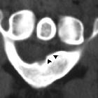
entrapment syndromes of the upper limb: a pictorial review. Causes of median nerve entrapment are illustrated. a: 56-year-old male with supracondylar spur seen on lateral elbow X-ray (white arrow). This is a known risk factor for median nerve impingement at the ligament of Struthers. b: Axial PD of 50-year-old female with carpal tunnel syndrome proven on electrophysiology. There is a congenital variant of bifid median nerve (white arrows) with persistent median artery (blue arrow) which can predispose to carpal tunnel syndrome. c: 30-year-old female with carpal tunnel syndrome. This patient has a ganglion (blue arrow) that is encroaching on the carpal tunnel. There is indentation of the median nerve with a width/height ratio of more than 3 (white arrow)

Nerve
entrapment syndromes of the upper limb: a pictorial review. Ultrasound thumb in patient with right thumb pain post trigger finger release. There is fusiform enlargement of the digital nerve (blue arrows) palmar-radial to the site of the prior A1 pulley release, extending over a length of 5 mm. Sonographic palpation elicited the usual symptom of pain radiating down the thumb. There is an echogenic focus (white arrow) immediately superficial to the nerve which may represent suture material (or calcification). This is consistent with entrapment given the proximity to the site of prior intervention

Nerve
entrapment syndromes of the upper limb: a pictorial review. Axial PD (a) and PDFS (b) palm of an 80-year-old female. There is increased oedema and volume loss in the intrinsic muscles of the hand radially (white arrows). This is consistent with deep ulnar nerve denervation pattern

Nerve
entrapment syndromes of the upper limb: a pictorial review. Cases of ulnar nerve entrapment at the elbow are illustrated. a Axial PDFS elbow in 50-year-old female with ulnar nerve neuropathy. The ulnar nerve (white arrow) returns increased signal intensity at the level of the cubital tunnel and is focally enlarged. b Sagittal PDFS elbow of a 54-year-old male with ulnar neuropathy. The ulnar nerve is enlarged (white arrow), with compression as it enters between the two flexor carpi ulnaris heads (blue arrows). c Axial PD elbow of a 60-year-old male showing the normal variant anconeus epitrochlearis (blue arrow) superficial to the ulnar nerve (white arrow) at the cubital tunnel. This can predispose to ulnar nerve entrapment

Nerve
entrapment syndromes of the upper limb: a pictorial review. The ulnar nerve is shown. The most common entrapment neuropathies of the ulnar nerve are the cubital tunnel syndrome at the elbow, and the ulnar tunnel syndrome at the wrist

Nerve
entrapment syndromes of the upper limb: a pictorial review. 50-year-old male with flexor pollicis longus palsy. Axial PDFS (a) and PD (b) at the level of the distal forearm demonstrate atrophy and denervation oedema of the pronator quadratus (white arrows), supportive of anterior interosseous nerve (AIN) denervation. A discrete lesion or compressive cause has not been identified

Nerve
entrapment syndromes of the upper limb: a pictorial review. Features and management of carpal tunnel syndrome on ultrasound are illustrated. a, b: 35-year-old female with carpal tunnel syndrome. The typical ultrasound appearance for neuropathy includes hypoechoic enlargement and nerve swelling (a, white arrow). There is an incidental variant of the recurrent branch of the median nerve that pierces the transverse carpal ligament (b, white arrow). c: Perineural cortisone injection of the median nerve under ultrasound guidance is shown. The needle (blue arrow) is positioned superficial and to the right side of the nerve (white arrow), with injectate encircling the nerve

Nerve
entrapment syndromes of the upper limb: a pictorial review. The median nerve is shown. Entrapment neuropathies of the median nerve include carpal tunnel syndrome, pronator teres syndrome and supracondylar process syndrome

Nerve
entrapment syndromes of the upper limb: a pictorial review. 37-year-old female with lateral scapular winging after schwannoma excision. Axial T1 at the level of the thoracic inlet (a) demonstrates asymmetry of the trapezius muscles (white arrows) with right-sided volume loss. Axial T1 fat-saturated post-contrast study (b) shows the avidly-enhancing schwannoma, which was located at level 2B in the upper posterior neck, deep to the upper sternocleidomastoid muscle and immediately posterior to the right internal jugular vein (blue arrow). This is along the expected path of the accessory nerve. While the nerve lesion in this case was caused by surgery, the illustrated muscle changes are also seen in entrapment

Nerve
entrapment syndromes of the upper limb: a pictorial review. PDFS (a) and PD sequence (b) of 40-year-old male with anconeus denervation oedema (white arrows) due to radial nerve neuropathy. The anconeus is supplied by the radial nerve before it bifurcates into the deep motor and superficial sensory branches, hence the cause of neuropathy in this case will be proximal to the bifurcation

Nerve
entrapment syndromes of the upper limb: a pictorial review. Cases of posterior interosseous nerve (PIN) pathology are illustrated: a, b Forearm ultrasound of a 58-year-old female who presented with arm pain. There is focal thickening throughout the right PIN (a, white arrows). This extends into the arcade of Frohse. This appearance can be seen with PIN syndrome. No adjacent collection or mass is identified. There was focal tenderness with transducer pressure. The left PIN is provided for comparison (b). c–e 60-year-old male who presented with progressive weakness of finger extension. Intramuscular lipoma of the supinator (blue arrow) resulted in PIN impingement at the arcade of Frohse (c). Axial T1 (c) and Axial PDFS images at the level of narrowing (d) and more distally (e), show focal thickening and hyperintensity of the PIN (c, d, white arrows). There is denervation oedema and atrophy of the extensor muscles (d, e, yellow arrows). f, g Axial PD (f) and PDFS (g) sequences with denervation oedema and atrophy of extensor muscles (white arrows) innervated by the PIN (not shown)

Nerve
entrapment syndromes of the upper limb: a pictorial review. Stack of PD axials of the upper forearm of a 55-year-old male post distal biceps tendon repair. The biceps tendon (a, b, c, white arrows) is intact although thickened just proximal to the tunnel entry. The radial nerve above the elbow appears normal (a, blue arrow). The superficial branch (b, yellow arrow) comes close to scarring around the enlarged biceps tendon and appears adherent. The deep branch (c, orange arrow) continues without abnormality to the supinator

Nerve
entrapment syndromes of the upper limb: a pictorial review. The radial nerve is shown. The most common entrapment neuropathy of the radial nerve is of the deep branch as it traverses the arcade of Frohse

Nerve
entrapment syndromes of the upper limb: a pictorial review. 49-year-old male with right upper limb weakness. Sagittal PD (a) shows moderate-severe fatty atrophy of the supraspinatus (white arrow) and infraspinatus muscles (blue arrow) which is compatible with suprascapular nerve entrapment. Interestingly, the site of the impingement is not in the typical suprascapular groove, but more proximally, secondary to ossification along the coracoclavicular ligament which is seen on sagittal CT (b, yellow arrow). The patient had a history of acromioclavicular sprain. Consecutive axial PD images (c, d) show that the ossification (c, yellow arrow) contacted and displaced the suprascapular vessels and nerve (c, d, orange arrows) posteromedially, stretching the nerve

Nerve
entrapment syndromes of the upper limb: a pictorial review. 20-year-old male with 6 months’ history of shoulder weakness. Axial (a) and sagittal (b) PDFS sequences show a posterior labral tear with paralabral cyst (white arrows) extending into the spinoglenoid notch. There is atrophy and mild denervation oedema in the infraspinatus muscle (b, blue arrow), sparing the supraspinatus since the site of entrapment is distal to the supraspinatus innervation

Nerve
entrapment syndromes of the upper limb: a pictorial review. 70-year-old with underwent MRI for rotator cuff tendinopathy. Sagittal proton density (PD) shoulder (a) shows severe fatty atrophy of the teres minor (white arrow). There is a lipomatous tumour with complete fat-suppression on the coronal PDFS sequence (b, yellow arrow), which is within the subscapularis muscle belly inferiorly and is impinging on the quadrilateral space and the traversing axillary nerve (a, blue arrow). This is consistent with chronic quadrilateral space syndrome

Nerve
entrapment syndromes of the upper limb: a pictorial review. Axial T1 and PDFS of a 70-year-old male with non-small cell lung cancer. There is a lytic destructive lesion in the mid-humeral shaft with cortical disruption and extraosseous extension (white arrow) extending into the deltoid attachment and the coracobrachialis muscle. While the musculocutaneous nerve is not confidently traced due to the motion artefact, it is expected to be in close contact with the lesion. Oedema signal within coracobrachialis muscle (yellow circle) can be either due to direct muscle involvement or denervation
Nervenkompressionssyndrome der oberen Extremität
Siehe auch:

 Assoziationen und Differentialdiagnosen zu Nervenkompressionssyndrome der oberen Extremität:
Kompressionssyndrom
des Nervus radialis


