cerebral oedema

School ager
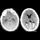
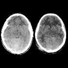
in a motor vehicle accidentAxial CT without contrast of the brain shows diffuse low density through the bilateral cerebral hemispheres (anteriorly) with a normal density cerebellum (posteriorly) that appears relatively hyperdense when compared to the cerebral density. Subarachnoid hemorrhage was also seen bilaterally in the basal cisterns.The diagnosis was diffuse cerebral edema.


Toddler who
is unresponsiveAxial CT without contrast of the brain shows high density material in the subgaleal tissues posteriorly, a wide lucency in the right posterior skull along with two areas of depressed lucency in the left frontal skull, a rounded high-density lesion in the midline of the cerebellum, and decreased density of the cerebrum when compared to the normal density of the cerebellum along with loss of the normal gray matter-white matter differentiation.The diagnosis was a posterior subgaleal hematoma, a right posterior diastatic skull fracture and a left frontal depressed skull fracture, cerebellar contusion and diffuse cerebral edema in a child abuse patient.

Infant who
has been shakenAxial CT without contrast of the brain shows normal density in the rounded top of the cerebellum (in the center of the left image) compared to the diffuse low-density throughout the cerebrum. There is obliteration of the basal cisterns and loss of the normal gray matter-white matter differentiation. There is also a small left sided high density extra-axial cresenteric fluid collection that tracks medially along the entire falx.The diagnosis was diffuse cerebral edema and a left-sided acute subdural hematoma with interhemispheric extension in a child abuse patient.

Cerebral
edema • Metastatic renal cell carcinoma to brain - Ganzer Fall bei Radiopaedia

Unconscious
school ager who fell from a tree. Axial CT with contrast of the brain shows diffuse brain swelling causing loss of the normal gyral pattern and compression of the ventricular system.The diagnosis was diffuse cerebral edema.

Toddler after
a motor vehicle accident. Axial CT without contrast of the brain shows acute hemorrhage around the Circle of Willis and brainstem (left) and in the posterior horns of the lateral ventricles bilaterally (right). There is also loss of the cortical sulci and basilar cisterns.The diagnosis was post-traumatic subarachnoid hemorrhage and intraventricular hemorrhage and diffuse cerebral edema.

Teenager in
motor vehicle accident with altered mental status. Axial CT without contrast of the brain obtained initially (left) shows loss of delineation of the cortical sulci and ventricular system. Axial CT without contrast obtained two days later (right) now also shows a round focus of hyperdensity at the right parietal gray matter white matter junction.The diagnosis was diffuse cerebral edema and delayed appearance of diffuse axonal injury.

Cerebral
edema • Combined cerebral edema - Ganzer Fall bei Radiopaedia

Cerebral
edema • Osmotic cerebral edema - Ganzer Fall bei Radiopaedia

Cerebral
edema • Late hyperacute ischemic stroke - Ganzer Fall bei Radiopaedia

Preschooler
after cardiac arrest. Axial CT without contrast of the head (above) shows a round mixed density lesion just to the right of the third ventricle along with diffuse subarachnoid hemorrhage and cerebral edema. Axial (below left) and sagittal (below right) CT angiogram shows the lesion to arise from the A1 segment of the right anterior cerebral artery.The diagnosis was giant intracranial aneurysm which had ruptured causing subarachnoid hemorrhage and diffuse cerebral edema.
Cerebral edema refers to a number of interconnected processes which result in abnormal shifts of water in various compartments of the brain parenchyma.
It has traditionally been broadly divided into vasogenic cerebral edema and cytotoxic cerebral edema, the latter a term commonly used to denote both true cytotoxic edema and ionic edema . In addition, although traditionally not included in discussions on edema, hemorrhagic transformation can be thought of as an extreme end-stage form of the same processes which lead to edema.
As such a more precise classification is :
Special types of edema to be considered:
- transependymal edema (also known as interstitial cerebral edema)
- combined cerebral edema
Siehe auch:
- Hirnabszess
- Pseudosubarachnoidalblutung
- Einklemmung Foramen magnum
- Hirntumoren
- Bluthirnschranke
- Höhenhirnödem
- causes of unilateral cerebral edema
und weiter:

 Assoziationen und Differentialdiagnosen zu Hirnödem:


