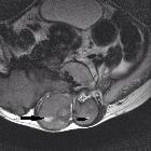
präsakrale Tumoren

Presacral
plexiform neurofibroma with urinary retention. Transverse ultrasound, pelvis. Multiple lobulated hypoechoic masses (white arrows) surround the rectum (red arrow). The rectal mucosa has normal bowel signature with no evidence of inflammation. The bladder is displaced anteriorly.

Presacral
plexiform neurofibroma with urinary retention. Coronal T2 Single Shot Fast Spin Echo (SSFE), abdomen and pelvis. Large, bulky mass anterior to the lower lumbar/upper sacral spine. Multiple internal hypointense tubular structures (target sign) with rim of hyperintensity (white arrows).

Presacral
plexiform neurofibroma with urinary retention. Axial T2 Fat-saturated Single Shot Fast Spin Echo (SSFE) through the pelvis demonstrates the large pelvic mass (white arrow) displacing the rectum and bladder anteriorly. A Foley catheter is in place (red arrow).

Presacral
plexiform neurofibroma with urinary retention. Sagittal T2 Fat-saturated SSFE through the pelvis. The large presacral mass (white arrow) shows involvement of the adjacent sacral nerve roots (red arrow) and associated widening of the sacral neural foramen.

Laparoscopically
excised retroperitoneal presacral Schwannoma: atypical pre and postoperative manifestations – case report. CT of the sagittal and coronal planes of the presacral region, respectively. Note the schwannoma (red circle), with dimensions of 4.4 × 3.9 × 3.4 cm

Toddler with
opsoclonus and myoclonus. Axial CT with contrast of the pelvis (above) shows (from top to bottom in the midline) contrast in the base of the bladder, contrast in the rectum which is deviated to the right, and a solid soft tissue mass anterior to the sacrum. Axial (below left) and sagittal (below right) T2 MRI without contrast of the pelvis shows the presacral mass to have high signal intensity.The diagnosis was presacral neuroblastoma.

An
interesting diagnosis for a presacral mass: case report. CT transverse view of lesion showing 33.52 mm diameter lesion in the presacral area.

An
interesting diagnosis for a presacral mass: case report. MRI Sagittal view of lesion.

An
interesting diagnosis for a presacral mass: case report. CT guided biopsy through presacral lesion using posterolateral approach.
präsakrale Tumoren
Siehe auch:
- Chordom
- Tumoren Os sacrum
- Riesenzelltumor des Sakrums
- Schwanzdarmzyste
- kindlicher präsakraler Tumor
- präsakrales Myelolipom
- präsakraler fetthaltiger Tumor
- solitäres sakrales Plasmozytom
- präsakrale extramedulläre Hämatopoese
- rectal carcinoma recurrence
- präsakrales Ganglioneurom
- präsakrales Liposarkom
- präsakrales Schwannom
- retrorektale zystische Entwicklungsstörungen
und weiter:

 Assoziationen und Differentialdiagnosen zu präsakrale Tumoren:


