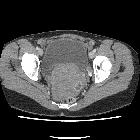
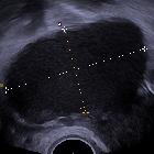
hemoperitoneum

Hemoperitoneum
• Ruptured corpus luteum cyst - Ganzer Fall bei Radiopaedia

Stumpfes
Bauchtrauma mit mesokolischer Einblutung rechts: Links oben hyperdenser Flüssigkeitssaum um die Leber und um die Milz, links Mitte fokale und diffuse Einblutung im rechten Mesocolon, links unten sagittal und rechts oben coronar beide Befunde. Rechts unten intraoperativer Situs mit großem Hämatom im Mesocolon.

School ager
gymnast who fell onto a balance beam. Axial CT with contrast of the abdomen (above) shows a low-density laceration in the center of the spleen with a round focus of high intensity in the middle of it felt to represent active bleeding. Axial CT with contrast of the pelvis (below) shows a large amount of fluid in the pelvis posterior to the bladder.The diagnosis was splenic trauma with active splenic bleeding and a large amount of hemoperitoneum.

Imaging for
acute pelvic pain in pregnancy. A 27-year-old woman presenting at the emergency department with acute pelvic pain. Abdominal US (a) shows a rounded lesion adjacent to the uterus, clearly separate from the left ovary. Axial CT scan shows a voluminous mass in the pelvic cavity (b); for the characterisation an MRI was required. Axial (c) and coronal (d) T2-weighted and T1-weighted images (e) of the pelvis show a right heterogeneous adnexal mass (arrow) with fallopian tube haematoma. Note the normal ovary (short arrow) in (c). Pre-contrast T1-weighted fat-saturated image (f) shows bloody ascites (short arrows). These findings are due to ectopic pregnancy with tubal rupture and haemoperitoneum

Ruptured
endometrioma in a nonpregnant patient: a case report. Large left pelvic hypodense structure (yellow arrow) drained by left gonadal vein (red arrow). There is moderate-volume ascites (blue arrows)

Ruptured
endometrioma in a nonpregnant patient: a case report. Hemoperitonieum with layering hyperdense material (blue arrow) compatible with blood products

Ruptured
endometrioma in a nonpregnant patient: a case report. Focal rim discontinuity of the dome of the left adnexal cystic mass (red arrow)

Hemoperitoneum
• Hemoperitoneum - Ganzer Fall bei Radiopaedia

Hemoperitoneum
• Acute mesenteric hematoma with active bleeding - Ganzer Fall bei Radiopaedia

Hemoperitoneum
• Hemoperitoneum post traumatic splenic injury - Ganzer Fall bei Radiopaedia

Hemoperitoneum
• Ruptured corpus luteum with large hemoperitoneum - Ganzer Fall bei Radiopaedia

Postoperative
bleeding after pancreaticoduodenectomy: CT findings and role. Multiplanar images from preoperative CT showed moderately dilated intrahepatic bile ducts (+), overdistended gallbladder (§), common bile duct dilatation (short arrows) with distal \"tapering\" stricture (arrowheads). Signs of vascular invasion and distant metastases were not seen.

Hemoperitoneum
• Ruptured ectopic pregnancy - Ganzer Fall bei Radiopaedia

Hemoperitoneum
• Hepatocellular carcinoma rupture with hemoperitoneum - Ganzer Fall bei Radiopaedia

Hemoperitoneum
• Ruptured corpus luteum cyst - Ganzer Fall bei Radiopaedia

Hemoperitoneum
• Ruptured ectopic pregnancy - Ganzer Fall bei Radiopaedia

Hemoperitoneum
• Ruptured corpus luteum cyst with hemoperitoneum - Ganzer Fall bei Radiopaedia

School ager
after motor vehicle accident with abdominal pain. Axial CT with contrast of the abdomen shows the bladder in the center of the image partially filled with contrast and surrounded by a large amount of free fluid throughout the pelvis.The diagnosis was splenic trauma with an extensive amount of hemoperitoneum.

Hemoperitoneum
• Ruptured corpus luteal cyst - Ganzer Fall bei Radiopaedia

Sheathed
goring: an unusual mechanism of trauma. MDCT revealed the presence of high-attenuated peritoneal fluid in the pelvic cavity (45 HU) due to haemoperitoneum.

Hemoperitoneum
• Polytrauma with multiple injuries - Ganzer Fall bei Radiopaedia
Hemoperitoneum (plural: haemoperitoneums) is the presence of blood within the peritoneal cavity.
Pathology
Etiology
- penetrating or non-penetrating abdominal trauma (often with associated organ injury)
- ruptured ectopic pregnancy
- ovarian cyst rupture
- aneurysm or pseudoaneurysm rupture
- neoplasm rupture
- acute hemorrhagic pancreatitis
- iatrogenic
- spontaneous bleeding, especially patients with coagulopathy or on anticoagulant therapy (uncommon)
Radiographic features
Ultrasound
- non-specific appearance of intraperitoneal free fluid
- may be hypo-, iso- or hyperechoic
- may demonstrate fluid-fluid levels with mixed internal echogenicity
CT
The density of fluid in the abdomen suggests its composition (i.e. ascites/bowel contents/bile vs hemorrhage) :
- recent hemorrhage (acute bleed) measures 30-45 HU
- clotted blood measures 45-70 HU
- old blood products/seroma or blood in patients with anemia may measure <30 HU
A collection of blood products can be homogeneous or heterogeneous (often low density with internal linear/nodular hyperdensities) ; fluid-fluid levels are often present.
MRI
- acute (<48 hours) hemoperitoneum has non-specific signal characteristics
- subacute (>3 weeks) may demonstrate concentric ring sign
- fluid-fluid levels (hematocrit effect) with high T1 / low T2 signal noted dependently
Differential diagnosis
- low-density hemoperitoneum may be indistinguishable from ascites or other causes of peritoneal free fluid
- there may be a delayed increase in ascites attenuation after administration of IV contrast (up to 3 days)
- there may be an increase of up to 25 HU
Siehe auch:
- Endometriose
- peritoneal cavity
- eitrige Perikarditis
- hämorrhagische Pankreatitis
- intraabdominelle Blutungen
und weiter:

 Assoziationen und Differentialdiagnosen zu Haemoperitoneum:


