tuberculous pelvic inflammatory disease

Extrapulmonary
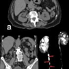
tuberculosıs: an old but resurgent problem. Sagittal-reformatted abdomen CT image of a 22-year-old female with lower abdominal pain and fever obtained after C/S surgery. The uterus is enlarged due to prior pregnancy. Free fluid is noted in the pelvis (arrows). Tuberculous pelvic inflammatory disease

Tuberculous
pelvic inflammatory disease • Genital tuberculosis - Ganzer Fall bei Radiopaedia

Tuberculous
pelvic inflammatory disease • Tubal involvement in genital tuberculosis - Ganzer Fall bei Radiopaedia

Female
genital tuberculosis. Multiplanar post-contrast images showed bilateral adnexal enlargement (+) with hypoattenuating (20-24 Hounsfield units, HU) content and uniform peripheral enhancement. No ascites and adenopathies were present.

Female
genital tuberculosis. Bilateral adnexal enlargement (+) with hypoattenuating (20-24 HU) content and peripheral enhancement. Dilated uterine cavity (*) by similar hypoattenuating content with thin endometrial enhancement (thin arrows).

Female
genital tuberculosis. Additionally, omental hazy infiltrate (arrowhead) was present. Note thin endometrial enhancement (thin arrows), 4 cm right-sided adnexal enlargement (+) with hypoattenuating (20-24 HU) content and peripheral enhancement.
Tuberculous pelvic inflammatory disease refers to pelvic inflammatory disease due to Mycobacterium tuberculosis.
Epidemiology
Genital tract involvement may be present in ~1.5% of cases of those affected with tuberculosis .
Pathology
Infection almost always results from spread from an extragenital source , usually from a haematogenous source or less commonly, via lymphatic vessels or from the peritoneal cavity.
Location
In the vast majority of cases, it involves the Fallopian tubes: tubal tuberculosis , Involvement is often bilateral .
Radiographic features
Hysterosalpingography (HSG)
- tubal involvement:
- obstruction of the Fallopian tube in the zone of transition between the isthmus and the ampulla
- multiple constrictions along the course of the Fallopian tube (resulting in a beaded appearance to the tube)
- endometrial involvement: features may vary; the spectrum according to one study was
- normal uterine cavity: ~57%
- irregular cavity: ~18.5%
- irregular filling defect: ~18.5%
- uterine synechiae: ~17%
- shrunken cavity: ~3%
- adnexal involvement
- they may be calcified lymph nodes or smaller, irregular calcifications in the adnexal area
CT
Tuberculous pelvic inflammatory disease may be associated with
- peritoneal involvement of tuberculosis (present in up to 50%)
- complex ascites
- thickened and nodular peritoneum
- lymphadenopathy
- necrotic
- calcified (chronic disease)
Treatment and prognosis
Complications
- infertility
- formation of tubo-ovarian abscesses
- tuberculous peritonitis: may be present in ~50% of cases
Differential diagnosis
For the hysterosalpingography appearance consider:
- salpingitis isthmica nodosa (SIN)
- Asherman syndrome: multiple adhesions
Siehe auch:
und weiter:


 Assoziationen und Differentialdiagnosen zu pelvine Tuberkulose: