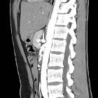
Truncus-coeliacus-Kompressionssyndrom

An unusual
cause of chronic abdominal pain: Median arcuate ligament syndrome. Axial image shows a filling defect on the proximal coeliac artery. Hypodense band surrounding the coeliac axis origin representing the median arcuate ligament (*).

Vascular
compression disorders • Median arcuate ligament syndrome - Ganzer Fall bei Radiopaedia

Chronic
mesenteric ischaemia due to median arcuate ligament syndrome. Green: coeliac trunk. Pink: superior mesenteric artery. Yellow: dilated duodeno-pancreatic arcades. Blue: common hepatic artery. Red: splenic artery.

Chronic
mesenteric ischaemia due to median arcuate ligament syndrome. White arrow: splenic artery

Chronic
mesenteric ischaemia due to median arcuate ligament syndrome. Blue arrow: proper hepatic artery

Chronic
mesenteric ischaemia due to median arcuate ligament syndrome. Red arrow: superior mesenteric artery. Green arrows: duodeno-pancreatic arcades

Chronic
mesenteric ischaemia due to median arcuate ligament syndrome. The catheter is in the abdominal aorta, and the extremity is at the ostium of the superior mesenteric artery.

Chronic
mesenteric ischaemia due to median arcuate ligament syndrome. Expirium. Please note the accentuation of the stenosis of the coeliac trunk by the median arcuate ligament, and also the increased post-stenotic dilatation.

Chronic
mesenteric ischaemia due to median arcuate ligament syndrome. Inspirium. About the same aspect of the coeliac trunk as in the CT examination.

Chronic
mesenteric ischaemia due to median arcuate ligament syndrome. Superior extrinsic stenosis of the first centimeters of the coeliac trunk, followed by a dilatation. Normal superior mesenteric artery.

Celiac artery
compression syndrome • Celiac artery compression syndrome - Ganzer Fall bei Radiopaedia

Celiac artery
compression syndrome • Median arcuate ligament syndrome (Dunbar syndrome) - Ganzer Fall bei Radiopaedia

Celiac artery
compression syndrome • Celiac artery compression syndrome - Ganzer Fall bei Radiopaedia

Celiac artery
compression syndrome • Celiac artery compression syndrome - Ganzer Fall bei Radiopaedia

CT-Angiographie
(parasagittale CPR) eines Truncus-coeliacus-Kompressionssyndroms

Celiac artery
compression syndrome • Median arcuate ligament syndrome - Ganzer Fall bei Radiopaedia

Celiac artery
compression syndrome • Median arcuate ligament syndrome (Dunbar syndrome) - Ganzer Fall bei Radiopaedia

Celiac artery
compression syndrome • Median arcuate ligament syndrome - Ganzer Fall bei Radiopaedia

Celiac artery
compression syndrome • Celiac artery compression by the diaphragmatic crurae - Ganzer Fall bei Radiopaedia

Celiac artery
compression syndrome • Median arcuate ligament syndrome - Ganzer Fall bei Radiopaedia

Celiac artery
compression syndrome • Median arcuate ligament syndrome - Ganzer Fall bei Radiopaedia

Celiac artery
compression syndrome • Median arcuate ligament syndrome and acute appendicitis - Ganzer Fall bei Radiopaedia

Celiac artery
compression syndrome • Median arcuate ligament syndrome - Ganzer Fall bei Radiopaedia

An unusual
cause of chronic abdominal pain: Median arcuate ligament syndrome. VR 3D image based on CT angiography data shows a significant stenosis of the coeliac artery in expiration.

An unusual
cause of chronic abdominal pain: Median arcuate ligament syndrome. Midline sagittal reformatted MIP image in deep inspiration (a) and expiration (b), revealing the increase of the degree of the proximal coeliac artery stenosis (up to 75% in expiration).

An unusual
cause of chronic abdominal pain: Median arcuate ligament syndrome. Aortic angio CT in deep expiration. Marked increase of the mild indentation of the MAL (*) on the proximal coeliac artery compared to the inspiration study, giving a “J-shape” or hooked appearance. Poststenotonic dilatation associated.

An unusual
cause of chronic abdominal pain: Median arcuate ligament syndrome. Sagittal arterial phase CT in deep inspiration shows focal narrowing on the superior aspect of the coeliac artery by the median arcuate ligament (*).

Teenager with
post prandial epigastric pain. Sagittal MIP (left) and 3D (right) CT with contrast of the abdomen in the venous phase obtained in expiration shows focal narrowing of the superior aspect of the proximal celiac trunk resulting in it having a hooked or J appearance with the absence of associated atherosclerosis.The diagnosis was median arcuate ligament syndrome.
Truncus-coeliacus-Kompressionssyndrom
Siehe auch:
- Arteria-mesenterica-superior-Syndrom
- Truncus coeliacus
- Hiatus aorticus
- vaskuläre Kompressionssyndrome
- Kompressionssyndrome
- diaphragmatic crurae
und weiter:

 Assoziationen und Differentialdiagnosen zu Truncus-coeliacus-Kompressionssyndrom:
Arteria-mesenterica-superior-Syndrom


