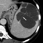
rupturierte Milzzyste
Laparoscopic
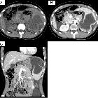
fenestration for a large ruptured splenic cyst combined with an elevated serum carbohydrate antigen 19–9 level: a case report. An abdominal CT scan showed a collapsed cystic lesion, measuring 12 cm × 12 cm × 8 cm, in the spleen and abdominal fluid in Morison’s pouch and around the liver and spleen (a). Moreover, an 8-mm cyst and a small collapsed cystic lesion were found posterior to the large cystic lesion on a contrast-enhanced CT scan (b). There was no evidence of contrast medium extravasation (c)

Laparoscopic
fenestration for a large ruptured splenic cyst combined with an elevated serum carbohydrate antigen 19–9 level: a case report. Contrast-enhanced MRI showed a cystic lesion, which exhibited slightly hyperintense signals on the T1- (a) and diffusion-weighted sequences and hyperintense signals on the T2-weighted sequence (b). No solid components or mural cysts were found in the cyst
rupturierte Milzzyste
Siehe auch:


 Assoziationen und Differentialdiagnosen zu rupturierte Milzzyste: