zystische Läsionen der Milz
Computed
tomography of the spleen: how to interpret the hypodense lesion. Transverse contrast-enhanced CT image acquired during the portal-venous phase in a 38-year-old woman illustrating a lymphangioma of the spleen. Note the lesion’s septations (arrow) that may enhance slightly after the administration of intravenous contrast material and homogeneous water-like content in the absence of solid components. The most important differential diagnosis to consider in this case would be a hydatid (echinococcal) cyst


Surgical
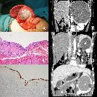
treatment of benign splenic lesions in pediatric patients: a case series of 30 cases from a single center. Splenic CECs (“*” marks the cysts). A A recurrent cyst after unroofing (Case 21). B The cyst compressing left renal and renal artery (Case 26). C Stratified squamous epithelium (HE staining). D Simultaneous separate CEC and hemagioma (white arrow shows the enhanced hemangioma, Case 19). E Cytokeratin immunohistochemical staining labels the epithelium. F Simultaneous separate CEC and omentum mesothelial cyst (white triangle shows the omentum mesothelial cyst, Case 28)


Zystische
Läsion der Milz mit relativ glatter, etwas unregelmäßiger, aber schmaler und verkalkter Begrenzung, am ehesten als z.B. posttraumatische Pseudozyste anzusehen.

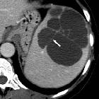
Computed
tomography of the spleen: how to interpret the hypodense lesion. Transverse contrast-enhanced CT images acquired during the portal-venous phase illustrating the different appearances of cystic splenic lesions. a A 73-year-old man with hydatid disease of the spleen (arrow). b A 23-year-old man with a congenital cyst of the spleen exhibiting water-like attenuation values. c A 52-year-old man with a multicystic metastasis from colon cancer (arrow). d A 63-year-old man with a false cyst, presumably after trauma (arrow)

Verkalkte
Pseudozyste der Milz, am ehesten posttraumatisch, jedoch nicht anamnestisch zu klären. Beachte auch die Sedimentation von röntgendichtem Material dorsal in der Pseudozyste.

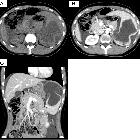
Teenager with
abdominal pain. Axial (above), coronal (below left) and sagittal (below right) CT with contrast of the abdomen shows a very large, thin walled, low density, non-septated lesion with undulating contours in the spleen.The diagnosis was a simple splenic cyst.
zystische Läsionen der Milz
Siehe auch:
- Milzhämangiom
- Pankreaspseudozyste
- verkalkte Milzzyste
- Wasserlilien-Zeichen
- Milzinfarkt
- Lymphangiom Milz
- septische Embolie Milz
- fokale Milzläsionen und Anomalien
- Splenic epidermoid cyst
- Echinokokkose der Milz
- Milzabszess
- Pseudozyste der Milz
- hypodense Milzläsionen
- rupturierte Milzzyste
- primary serous cyst of the spleen
und weiter:


 Assoziationen und Differentialdiagnosen zu zystische Läsionen der Milz: