splenic epidermoidcyst
Surgical
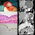
treatment of benign splenic lesions in pediatric patients: a case series of 30 cases from a single center. Splenic CECs (“*” marks the cysts). A A recurrent cyst after unroofing (Case 21). B The cyst compressing left renal and renal artery (Case 26). C Stratified squamous epithelium (HE staining). D Simultaneous separate CEC and hemagioma (white arrow shows the enhanced hemangioma, Case 19). E Cytokeratin immunohistochemical staining labels the epithelium. F Simultaneous separate CEC and omentum mesothelial cyst (white triangle shows the omentum mesothelial cyst, Case 28)

Spontaneous
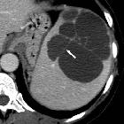
regression of a true splenic cyst: a case report and review of the literature. CT scan (1997): A 6 cm splenic cyst is evident at the lower pole of the spleen.

Spontaneous
regression of a true splenic cyst: a case report and review of the literature. CT scan (2005): A 3 × 5 cm splenic cyst is seen located at the lower splenic pole.

Spontaneous
regression of a true splenic cyst: a case report and review of the literature. MRI (2006): A 1.8 × 1.4 cm cystic lesion has remained with a low T2 sign in the peripheral border.

Massive
splenic cyst in pregnancy: case report. MRI of splenic cyst and fetus. a coronial view; b sagittal view

Subtotal
resection and omentoplasty of the epidermoid splenic cyst: a case report. CT scan image showing a well-defined cystic lesion on the upper part of the spleen.
Laparoscopic
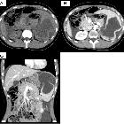
fenestration for a large ruptured splenic cyst combined with an elevated serum carbohydrate antigen 19–9 level: a case report. An abdominal CT scan showed a collapsed cystic lesion, measuring 12 cm × 12 cm × 8 cm, in the spleen and abdominal fluid in Morison’s pouch and around the liver and spleen (a). Moreover, an 8-mm cyst and a small collapsed cystic lesion were found posterior to the large cystic lesion on a contrast-enhanced CT scan (b). There was no evidence of contrast medium extravasation (c)

Laparoscopic
fenestration for a large ruptured splenic cyst combined with an elevated serum carbohydrate antigen 19–9 level: a case report. Contrast-enhanced MRI showed a cystic lesion, which exhibited slightly hyperintense signals on the T1- (a) and diffusion-weighted sequences and hyperintense signals on the T2-weighted sequence (b). No solid components or mural cysts were found in the cyst
splenic epidermoidcyst
Siehe auch:


 Assoziationen und Differentialdiagnosen zu splenic epidermoidcyst: