fokale Milzläsionen und Anomalien

Hämangiom
der Milz mit typischem Irisblendenphänomen: Oben arteriell mit randständiger Kontrastmittelaufnahme, unten portalvenös aufgefüllt. (nicht histologisch bestätigt, aber typisch)

Extrapulmonale
Sarkoidose bei einem 36jährigen Mann. Befall von Leber und Milz sowie der Lunge. Die Computertomographie axial zeigt multiple hypodense Herde, die primär an ein Malignom denken ließen, zumal die Befunde in der PET-CT stark stoffwechselaktiv waren.

Littoralzellangiome
der Milz in der Magnetresonanztomografie: Links T2 und T1, rechts Kontrastmitteldynamik.

Computed
tomography of the spleen: how to interpret the hypodense lesion. Transverse contrast-enhanced CT images acquired during the portal-venous phase. a A 54-year-old woman with a hamartoma that exhibits mild contrast enhancement. b A 76-year-old woman with a hamartoma that presents with focal areas of fat attenuation

Computed
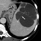
tomography of the spleen: how to interpret the hypodense lesion. Transverse contrast-enhanced CT image acquired during the portal-venous phase in a 38-year-old woman illustrating a lymphangioma of the spleen. Note the lesion’s septations (arrow) that may enhance slightly after the administration of intravenous contrast material and homogeneous water-like content in the absence of solid components. The most important differential diagnosis to consider in this case would be a hydatid (echinococcal) cyst

Hämangiom
der Milz in der Magnetresonanztomografie: Links T2 und T1, rechts Kontrastmitteldynamik mit Irisblendenphänomen.

Splenic
lesions and anomalies • B-cell lymphoma of the spleen - Ganzer Fall bei Radiopaedia

Splenunculus
• Splenunculus - Ganzer Fall bei Radiopaedia

Splenic
lesions and anomalies • Calcified splenic cyst - Ganzer Fall bei Radiopaedia

Splenic
lesions and anomalies • Postpartum splenic abscess - Ganzer Fall bei Radiopaedia

Splenic
lesions and anomalies • Hydatid cyst of the spleen - Ganzer Fall bei Radiopaedia

Splenosis •
Splenosis - Ganzer Fall bei Radiopaedia

Sickle cell
disease • Sickle cell disease - Ganzer Fall bei Radiopaedia

Splenic
hemangioma • Splenic hemangioma - Ganzer Fall bei Radiopaedia


Splenic
lesions and anomalies • B-cell lymphoma involving the spleen - Ganzer Fall bei Radiopaedia

Splenic
lesions and anomalies • Mantle cell lymphoma - Ganzer Fall bei Radiopaedia

Splenic cyst
• Splenic epidermoid cyst - Ganzer Fall bei Radiopaedia

Sclerosing
angiomatoid nodular transformation of the spleen • Sclerosing angiomatoid nodular transformation of the spleen (SANT) - Ganzer Fall bei Radiopaedia

Leishmaniasis
• Splenic leishmaniasis - Ganzer Fall bei Radiopaedia

Splenomegaly
• Splenic infarct - Ganzer Fall bei Radiopaedia

Splenic
lesions and anomalies • Bipartite spleen - Ganzer Fall bei Radiopaedia

Splenic
lesions and anomalies • Calcified tuberculous granulomas - Ganzer Fall bei Radiopaedia

Splenic
lesions and anomalies • Giant splenic pseudocyst - Ganzer Fall bei Radiopaedia

Sarkoidose
der Milz in der Computertomographie: Viele kleine Knötchen in der Milz rechts im Bild.

Computed
tomography of the spleen: how to interpret the hypodense lesion. Transverse contrast-enhanced CT images acquired during the late arterial phase. a A 23-year-old man who was involved in a motorcycle accident and suffered a splenic laceration with extensive intraparenchymal, subcapsular and perisplenic haematoma. b A 34-year-old woman who was involved in a motor vehicle accident and suffered a splenic laceration. Note the active contrast extravasation within the spleen (arrow)

Abszess in
der Milz bei Salmonella: Beachte auch die unscharfe Kontur der Begrenzung nach ventral.

Computed
tomography of the spleen: how to interpret the hypodense lesion. Transverse contrast-enhanced CT image acquired during the portal-venous phase in a 47-year-old woman with metastatic ovarian cancer. The newly diagnosed hypodense lesion within the spleen (arrow) needs to be regarded as a metastasis unless proven otherwise

Computed
tomography of the spleen: how to interpret the hypodense lesion. a, b Transverse contrast-enhanced CT images acquired during the portal-venous phase at two different levels in a 55-year-old man with diffuse large B-cell lymphoma exhibiting multiple hypodense, mildly contrast-enhancing lesions within the spleen (arrows); with the latter usually being enlarged

Computed
tomography of the spleen: how to interpret the hypodense lesion. a, b Transverse contrast-enhanced CT images acquired during the portal-venous phase at two different levels in a 55-year-old man with littoral cell angioma, which presents as multiple hypodense, partially contrast-enhancing, lesions of different size (arrows)

Computed
tomography of the spleen: how to interpret the hypodense lesion. a, b Transverse non-contrast-enhanced CT images acquired at two different levels in a 55-year-old woman with splenic peliosis, exhibiting multiple hypodense lesions of different size (arrows) within a massively enlarged spleen

Computed
tomography of the spleen: how to interpret the hypodense lesion. a, b Transverse contrast-enhanced CT images acquired during the portal-venous phase at different levels in a 55-year-old woman with sarcoidosis affecting the liver (arrowhead) and the spleen (short arrow, long arrows)

Computed
tomography of the spleen: how to interpret the hypodense lesion. Transverse contrast-enhanced CT images acquired during the portal-venous phase. a A 19-year-old woman with multiple pyogenic splenic abscesses during a period of immunosuppression and haematogenous spread of Staphylococcus aureus (short arrows). b A 48-year-old man with a pyogenic splenic abscess exhibiting gas formations and subcapsular fluid accumulation due to a spontaneous rupture of the abscess. c A 27-year-old man with multiple tuberculous abscesses (long arrows)

Computed
tomography of the spleen: how to interpret the hypodense lesion. Transverse contrast-enhanced CT images acquired during the portal-venous phase. a A 49-year-old woman with a large splenic infarction secondary to thromboembolism from atrial fibrillation. b A 42-year-old woman with a complete splenic infarction, partial renal infarction (arrow) and hepatic infarction (arrowhead) due to a cardiogenic shock caused by sudden cardiac arrest

Splenic
lesions and anomalies • Splenic lipoma - Ganzer Fall bei Radiopaedia

Computed
tomography of the spleen: how to interpret the hypodense lesion. Transverse contrast-enhanced CT images acquired during the portal venous phase. a A 43-year-old man with advanced sickle-cell disease. Note the irregular shape of the spleen, as well as the increased density of the splenic parenchyma, together with extensive calcifications as a consequence of constantly occurring micro-infarctions. b A 38-year-old man with sickle-cell disease exhibiting end-stage splenic involvement. Note the increased density, calcifications of the shrunken spleen

Computed
tomography of the spleen: how to interpret the hypodense lesion. Transverse CT images acquired (a) before and (b, c) after intravenous administration of iodinated contrast material in a 74-year-old woman with a splenic haemangioma. Note the typical nodular enhancement beginning in the arterial phase and extending in a centripetal manner during the portal-venous phase

Computed
tomography of the spleen: how to interpret the hypodense lesion. Transverse contrast-enhanced CT images acquired during the portal-venous phase illustrating the different appearances of cystic splenic lesions. a A 73-year-old man with hydatid disease of the spleen (arrow). b A 23-year-old man with a congenital cyst of the spleen exhibiting water-like attenuation values. c A 52-year-old man with a multicystic metastasis from colon cancer (arrow). d A 63-year-old man with a false cyst, presumably after trauma (arrow)

Computed
tomography of the spleen: how to interpret the hypodense lesion. Transverse CT images acquired (a) before and (b, c) after the intravenous administration of iodinated contrast material in a 38-year-old man. b Note the trabecular enhancement pattern of the spleen during the arterial phase, when compared with the homogeneous appearance of the spleen during the (c) portal venous phase and on the (a) non-enhanced image


Sclerosing
angiomatoid nodular transformation of the spleen mimicking metastasis of melanoma: a case report and review of the literature. a B-mode ultrasound picture of the lesion. b, c, d Contrast-enhanced ultrasound image 19, 72, and 152 seconds after injection of 1 ml SonoVue®

Sclerosing
angiomatoid nodular transformation of the spleen mimicking metastasis of melanoma: a case report and review of the literature. a Computed tomography scan of the abdomen showing two hypodense lesions of the spleen. b, c Magnetic resonance imaging of the abdomen showing the two lesions in T1 and T2 mode, respectively
There are a number of splenic lesions and anomalies:
Congenital anomalies
Mass lesions
Benign mass lesions
- splenic cyst (mnemonic)
- splenic pseudocyst
- splenic hemangioma: commonest benign splenic lesion
- splenic lymphangioma
- splenic hamartoma
- sclerosing angiomatoid nodular transformation (SANT): fibrosing variant of hamartoma
- extramedullary hematopoiesis in the spleen
- splenic abscess: sonographically characterized by multiple “target” lesions
- splenic hydatid cysts
- splenic inflammatory pseudotumor
- splenic lipoma
- splenic angiomyolipoma
- splenic fibroma
- sarcoidosis
- focal splenic lesions in type I Gaucher disease
- splenic hematoma
Indeterminate mass lesions
Malignant mass lesions
- splenic lymphoma: commonest malignant tumor with splenic involvement
- angiosarcoma of spleen: commonest primary malignant splenic tumor
- haemagiopericytoma of spleen
- splenic metastases: 50% of which are from malignant melanoma
- splenic malignant fibrous histocytoma
Diffuse infiltrative processes
These usually manifest as splenomegaly. Some can present as distinct lesions (e.g. granulomas).
Multifocal splenic lesions
- lymphoma
- metastases
- sarcoidosis
- fungal abscesses
- granulomatous infections
- gamna-gandy bodies
Other abnormalities
- splenic rupture
- traumatic: spleen is the most frequently injured intra-abdominal organ in blunt trauma
- non-traumatic
- splenic infarction
- splenosis
- splenic peliosis
Siehe auch:
- verkalkte Milzläsionen
- Raumforderungen der Milz
- Sarkoidose der Milz
- Milzhämangiom
- Splenomegalie
- zystische Läsionen der Milz
- Nebenmilz
- Splenose
- Milzinfarkt
- Angiosarkom der Milz
- Lymphom der Milz
- Milzmetastasen
- Lymphangiom Milz
- ektopes Milzgewebe
- Littoralzellangiom der Milz
- Milzhamartom
- Polysplenie-Syndrom
- Echinokokkose der Milz
- splenic epidermoidcyst
- Milzabszess
- hypodense Milzläsionen
- Hämangioperizytom der Milz
- sklerosierende angiomatöse noduläre Transformation der Milz
- splenic involvement leukemia
- haemagiopericytoma of spleen
- splenic siderosis
und weiter:
- hypointense Milzherde
- Differentialdiagnose eingeblutete Raumforderung Milz
- extramedulläre Hämatopoese in der Milz
- granulomatöse Erkrankungen der Milz
- echoreiche Milzläsion
- Differentialdiagnose Raumforderung Milz hypervaskularisiert
- arteriell anreichernder Milzherd
- splenic lesions in liver cirrhosis
- contrast-enhanced ultrasound for the evaluation of splenic infarctions

 Assoziationen und Differentialdiagnosen zu fokale Milzläsionen und Anomalien:
sklerosierende
angiomatöse noduläre Transformation der Milz


