Angiosarkom der Milz


Angiosarcomas of the spleen are rare malignant splenic neoplasms. The term is usually given to describe a primary angiosarcoma of the spleen although angiosarcoma elsewhere can also rarely metastasize to the spleen. Despite its absolute rarity, a splenic angiosarcoma is considered the most common primary non-hematolymphoid splenic malignancy .
Epidemiology
The general consensus is that there is no recognized gender predilection (occurs almost equally in females and males). Occasional publications, however, suggest a slight male predilection . Commonly seen in older patients, with the peak incidence is thought to be around the 6 decade.
Clinical presentation
Clinical symptomatology can be highly variable, often posing difficult diagnostic problems.
Pathology
Macroscopic examination often shows splenomegaly with cut sections revealing discrete lesions in a majority of cases. These can range from well-circumscribed firm nodules to poorly delineated foci of necrosis and hemorrhage associated with cystic spaces.
Microscopically, the tumors are often heterogeneous. The lesions typically demonstrate focal vasoformative component lined by atypical endothelial cells. Solid sarcomatous, papillary, and epithelioid growth patterns can be observed.
Associations
Unlike with primary hepatic angiosarcoma, there is no known association between splenic angiosarcoma and occupational exposure to chemicals, such as vinyl chloride or arsenic, or prior injection with the contrast agent thorium dioxide .
Radiographic features
Ultrasound
Reported sonographic features include splenomegaly with largely heterogeneous echotexture, including cystic and solid components . Increased vascularity on Doppler is commonly present .
CT
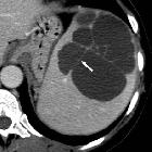
CT may show solitary or multiple nodular masses of heterogeneous low attenuation in an enlarged spleen; necrosis and hemorrhagic areas may account for the heterogeneity . There is generally irregular and poorly defined contours .
Occasional large subcapsular intrasplenic or perisplenic extracapsular blood collections (hemoperitoneum) may be present.
Some of these masses may show peripheral enhancement with the margins of the lesions often irregular or poorly defined.
Less frequently, CT scans may show a moderate splenomegaly with a micronodular involvement of the organ .
- pre-contrast CT: the tumors may appear hyperattenuating due to components of acute hemorrhage
- dynamic contrast-enhanced CT scans: the lesions may exhibit substantial peripheral contrast enhancement similar to that of hepatic hemangiomas
MRI
Reported MRI features include:
- T1 and T2:
- nodular hypointense (relative to the normal adjacent splenic parenchyma) masses on both T1- and T2-weighted images
- large masses with increased signal intensity on both T1- and T2-weighted images that are likely related to areas of subacute hemorrhage, as well as tumor necrosis
- areas of decreased signal intensity within the tumor, owing to chronic hemorrhage with hemosiderin deposition
- C+ (Gd): usually shows intense and multinodular (heterogenous ) enhancement with focal areas of non-enhancement, likely representing intratumoral hemorrhage and necrosis
Treatment and prognosis
It is an extremely aggressive fatal neoplasm at is almost universally fatal (median survival at approximately 24-36 months at the time of initial writing) despite treatment . Distant metastases occur most frequently in the liver (approximately 70% of cases), lung, pleural lymph nodes, bone, and brain. Prompt splenectomy prior to splenic rupture may improve survival .
Complications
- spontaneous splenic rupture : can occur in up to 30% of cases
History and etymology
It was first described in 1879 by T Langhans .
Differential diagnosis
General imaging differential considerations include vascular splenic lesions such as:
- splenic hemangioma
- Littoral cell angioma of spleen
- lymphangioma of spleen
- hemangiopericytoma of spleen
See also
- angiosarcoma - general
- splenic lesions and anomalies
Siehe auch:
- Milzhämangiom
- Angiosarkom
- Lymphangiom Milz
- Littoralzellangiom der Milz
- fokale Milzläsionen und Anomalien
- Hämangioperizytom der Milz
und weiter:


 Assoziationen und Differentialdiagnosen zu Angiosarkom der Milz: