hypodense Milzläsionen

Computed
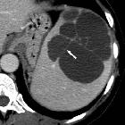
tomography of the spleen: how to interpret the hypodense lesion. Transverse contrast-enhanced CT image acquired during the portal-venous phase in a 38-year-old woman illustrating a lymphangioma of the spleen. Note the lesion’s septations (arrow) that may enhance slightly after the administration of intravenous contrast material and homogeneous water-like content in the absence of solid components. The most important differential diagnosis to consider in this case would be a hydatid (echinococcal) cyst

Computed
tomography of the spleen: how to interpret the hypodense lesion. Transverse contrast-enhanced CT images acquired during the portal-venous phase. a A 54-year-old woman with a hamartoma that exhibits mild contrast enhancement. b A 76-year-old woman with a hamartoma that presents with focal areas of fat attenuation

Computed
tomography of the spleen: how to interpret the hypodense lesion. Transverse CT images acquired (a) before and (b, c) after the intravenous administration of iodinated contrast material in a 38-year-old man. b Note the trabecular enhancement pattern of the spleen during the arterial phase, when compared with the homogeneous appearance of the spleen during the (c) portal venous phase and on the (a) non-enhanced image

Computed
tomography of the spleen: how to interpret the hypodense lesion. Transverse contrast-enhanced CT images acquired during the portal-venous phase illustrating the different appearances of cystic splenic lesions. a A 73-year-old man with hydatid disease of the spleen (arrow). b A 23-year-old man with a congenital cyst of the spleen exhibiting water-like attenuation values. c A 52-year-old man with a multicystic metastasis from colon cancer (arrow). d A 63-year-old man with a false cyst, presumably after trauma (arrow)

Computed
tomography of the spleen: how to interpret the hypodense lesion. Transverse contrast-enhanced CT images acquired during the late arterial phase. a A 23-year-old man who was involved in a motorcycle accident and suffered a splenic laceration with extensive intraparenchymal, subcapsular and perisplenic haematoma. b A 34-year-old woman who was involved in a motor vehicle accident and suffered a splenic laceration. Note the active contrast extravasation within the spleen (arrow)

Computed
tomography of the spleen: how to interpret the hypodense lesion. Transverse contrast-enhanced CT images acquired during the portal-venous phase. a A 19-year-old woman with multiple pyogenic splenic abscesses during a period of immunosuppression and haematogenous spread of Staphylococcus aureus (short arrows). b A 48-year-old man with a pyogenic splenic abscess exhibiting gas formations and subcapsular fluid accumulation due to a spontaneous rupture of the abscess. c A 27-year-old man with multiple tuberculous abscesses (long arrows)

Computed
tomography of the spleen: how to interpret the hypodense lesion. Transverse contrast-enhanced CT images acquired during the portal venous phase. a A 43-year-old man with advanced sickle-cell disease. Note the irregular shape of the spleen, as well as the increased density of the splenic parenchyma, together with extensive calcifications as a consequence of constantly occurring micro-infarctions. b A 38-year-old man with sickle-cell disease exhibiting end-stage splenic involvement. Note the increased density, calcifications of the shrunken spleen

Computed
tomography of the spleen: how to interpret the hypodense lesion. Transverse contrast-enhanced CT images acquired during the portal-venous phase. a A 49-year-old woman with a large splenic infarction secondary to thromboembolism from atrial fibrillation. b A 42-year-old woman with a complete splenic infarction, partial renal infarction (arrow) and hepatic infarction (arrowhead) due to a cardiogenic shock caused by sudden cardiac arrest

Computed
tomography of the spleen: how to interpret the hypodense lesion. Transverse CT images acquired (a) before and (b, c) after intravenous administration of iodinated contrast material in a 74-year-old woman with a splenic haemangioma. Note the typical nodular enhancement beginning in the arterial phase and extending in a centripetal manner during the portal-venous phase

Computed
tomography of the spleen: how to interpret the hypodense lesion. a, b Transverse contrast-enhanced CT images acquired during the portal-venous phase at different levels in a 55-year-old woman with sarcoidosis affecting the liver (arrowhead) and the spleen (short arrow, long arrows)

Computed
tomography of the spleen: how to interpret the hypodense lesion. a, b Transverse non-contrast-enhanced CT images acquired at two different levels in a 55-year-old woman with splenic peliosis, exhibiting multiple hypodense lesions of different size (arrows) within a massively enlarged spleen

Computed
tomography of the spleen: how to interpret the hypodense lesion. a, b Transverse contrast-enhanced CT images acquired during the portal-venous phase at two different levels in a 55-year-old man with littoral cell angioma, which presents as multiple hypodense, partially contrast-enhancing, lesions of different size (arrows)

Computed
tomography of the spleen: how to interpret the hypodense lesion. a, b Transverse contrast-enhanced CT images acquired during the portal-venous phase at two different levels in a 55-year-old man with diffuse large B-cell lymphoma exhibiting multiple hypodense, mildly contrast-enhancing lesions within the spleen (arrows); with the latter usually being enlarged

Computed
tomography of the spleen: how to interpret the hypodense lesion. Transverse contrast-enhanced CT image acquired during the portal-venous phase in a 47-year-old woman with metastatic ovarian cancer. The newly diagnosed hypodense lesion within the spleen (arrow) needs to be regarded as a metastasis unless proven otherwise
 Assoziationen und Differentialdiagnosen zu hypodense Milzläsionen:


