splenic metastases

Milz- und
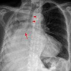
Lebermetastasen bei einem niedrig differenzierten Adenokarzinom des Sigmas. Computertomorgaphie axial und coronar.

Computed
tomography of the spleen: how to interpret the hypodense lesion. Transverse contrast-enhanced CT image acquired during the portal-venous phase in a 47-year-old woman with metastatic ovarian cancer. The newly diagnosed hypodense lesion within the spleen (arrow) needs to be regarded as a metastasis unless proven otherwise
Splenic metastases are relatively rare on imaging, although they are more commonly found on autopsy. Typically they are part of a widespread metastatic disease.
Epidemiology
The rate of splenic metastases varies between 1-10% of autopsy studies, depending on whether microscopic or macroscopic metastases were included .
Pathology
Most commonly metastases are a solitary splenic (solid or cystic) mass, may rarely be infiltrative . Primary sources include :
- cutaneous malignant melanoma (most common)
- breast cancer
- ovarian cancer
- colorectal cancer
- endometrial carcinoma
- gastric cancer
- lung cancer
- clear cell renal carcinoma
- pancreatic neuroendocrine tumor
- thymic carcinoma
- prostatic adenocarcinoma
- esophageal cancer
Radiographic features
Ultrasound
- solitary or multiple well-defined masses
- most commonly appear as a hypoechoic lesion (target appearance), although can be iso- or hyperechoic
- contrast-enhanced US (CEUS): rapid wash-in and wash-out
CT
- usually hypoattenuating masses
- cystic components may be present
MRI
- T1: low signal intensity
- T2: high signal intensity
- C+ (Gd): variable, mostly depending on the primary malignancy
Siehe auch:
- Lungenkarzinom
- Kolorektales Karzinom
- Neoplasien des Ovars
- Lymphom der Milz
- Malignes Melanom Metastase
- fokale Milzläsionen und Anomalien
- Neoplasien der Mamma
- granulomatöse Erkrankungen der Milz
und weiter:


 Assoziationen und Differentialdiagnosen zu Milzmetastasen: