colorectal carcinoma


Colorectal carcinoma (CRC) is the most common cancer of the gastrointestinal tract and the second most frequently diagnosed malignancy in adults. CT and MRI are the modalities most frequently used for staging. Surgical resection may be curative although five-year survival rate is 40-50%.
Epidemiology
Colorectal carcinoma is common, accounting for 15% of all newly diagnosed cancers, and tends to be a disease of the elderly, with the median age of diagnosis between 60 and 80 years of age , slightly younger for rectal carcinoma. There is also a slight male predilection for rectal cancers, not found in tumors elsewhere in the colon.
Risk factors
A number of predisposing factors have been identified, including:
- low fiber and high fat and animal protein diet
- obesity (especially in men)
- inflammatory bowel disease (IBD)
- chronic ulcerative colitis
- Crohn disease (particularly in bypassed loops/in vicinity of chronic fistula)
- asbestos workers
- a family history of benign/malignant colorectal tumors
- history of endometrial/breast cancer
- pelvic irradiation
- ureterosigmoidostomy
- colonic adenoma
- dysplasia of colon within flat mucosa
- prominent lymphoid follicular pattern
Associations
Syndromes
Recognized hereditary syndromes are seen in 6% of colorectal carcinomas. These include:
- familial adenomatous polyposis syndrome (FAP)
- Gardner syndrome variant
- Turcot syndrome variant
- Peutz-Jeghers syndrome
- hereditary non-polyposis colon cancer syndrome (HNPCC)
Clinical presentation
Clinical presentation is typically insidious:
- altered bowel habit (constipation and/or diarrhea)
- iron-deficiency anemia (chronic occult blood loss)
However initial manifestation may be acute:
- bowel obstruction
- intussusception
- heavy rectal bleeding
Occasionally metastatic disease may be the first sign.
Positive blood cultures or bacterial endocarditis with Streptococcus bovis is strongly suggestive of underlying colorectal cancer .
In general:
- right-sided tumors are larger and present with a mass, distant disease or iron deficiency anemia
- left-sided tumors present earlier with altered bowel habit
Pathology
Colorectal cancers, 98% of which are adenocarcinomas, arise in the vast majority of cases from pre-existing colonic adenomas (neoplastic polyps), which progressively undergo a malignant transformation as they accumulate additional mutations (so-called multihit hypothesis).
Morphologically cancers can be:
- sessile
- exophytic
- circumferential (apple core)
- ulcerated
- desmoplastic
Rarely the malignant cells will widely invade the submucosa, analogous to linitis plastic of the stomach. These are typically scirrhous adenocarcinomas (signet-ring type).
Metastases may be widespread in advanced disease, although the liver is by far the most common site involved.
Specific subtypes
Other usual types include
- primary colorectal small cell carcinoma: extremely rare
Location
Colorectal cancers can be found anywhere from the cecum to the rectum, in the following distribution :
- rectosigmoid: 55%
- cecum and ascending colon: ~20%
- ileocecal valve: 2%
- transverse colon: ~10%
- descending colon: ~5%
Staging
See: colon cancer staging
Radiographic features
Fluoroscopy
Barium enema
- sensitivities for polyps >1 cm
- single contrast: 77-94%
- double-contrast: 82-98%
- polyps <1 cm: <50% detection
Appearances will reflect macroscopic appearance, with lesions seen as filling defects. These need to be differentiated from residual fecal matter. Typically they appear as exophytic or sessile masses or maybe circumferential (apple core sign). Fistulas to bladder, vagina, or bowel may also be demonstrated.
Rarely the stenotic segment will be long particularly with scirrhous adenocarcinomas.
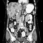
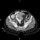
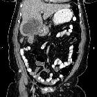
CT
CT is the modality most used for staging colorectal carcinoma, with an accuracy of only between 45-77% , able to assess nodes and metastases.
It is often able to diagnose tumors although it is insensitive to small masses. CT colonography is increasing in popularity as an alternative to colonoscopy.
Most colorectal carcinomas are of soft tissue density that narrow the bowel lumen . Ulceration in larger mass is also seen. Occasionally low-density masses with low-density lymph nodes are seen in mucinous adenocarcinoma, due to the majority of the tumor composed of extracellular mucin. Psammomatous calcifications in mucinous adenocarcinoma can also be present.
Complications may also be evident, e.g. fistulae, obstruction, intussusception, perforation .
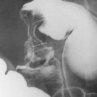
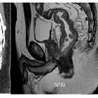
MRI
MRI has a staging accuracy of 73% with a 40% sensitivity for lymph node metastases . MRI is having an increasing role to play in the staging of rectal cancer.
Treatment and prognosis
Treatment involves local control with resection in almost all cases. Adjuvant chemotherapy is reserved for stage III disease.
Overall 5-year survival rate is 40-50%, with the stage at operation the single most important factor affecting prognosis.
- Duke A: 80-90%
- Duke B: 70%
- Duke C: 33%
- Duke D: 5%
Recurrence in common:
- local recurrence at the line of anastomosis: tend to occur within two years of diagnosis (80%)
- distant metastatic recurrence
The tumor marker CEA is routinely used for detecting postoperative early recurrence and metastatic disease (especially liver disease). It is also used for monitoring response to treatment of metastatic disease
- as with most tumor markers, it is inappropriate for screening given its poor sensitivity and specificity
- higher levels of CEA are associated with:
- higher-grade tumors
- higher-stage disease
- visceral metastases (especially liver metastases)
Screening recommendations
Screening recommendations are contentious and vary widely from country to country. An example would be:
- for persons >50 years of age: an annual fecal occult blood test (often a fecal immunochemical test (FIT)) and sigmoidoscopy/barium enema every 3 to 5 years
- for first-degree relatives of patients with colon cancer: screening should start at age 40
Differential diagnosis
General imaging differential considerations on CT include:


 Assoziationen und Differentialdiagnosen zu Kolorektales Karzinom:


