Gardner-Syndrom

Pharyngeal
desmoid-type fibromatosis in a pediatric patient with Gardner"s syndrome. Axial CT image shows an oropharyngeal well circumscribed, soft tissue mass, with similar attenuation than the muscles, and slight enhancement after contrast administration. The airway is almost completely occupied by the mass.

Pharyngeal
desmoid-type fibromatosis in a pediatric patient with Gardner"s syndrome. Coronal CT image depicts the same oropharyngeal soft tissue mass.

Pharyngeal
desmoid-type fibromatosis in a pediatric patient with Gardner"s syndrome. On T2-weighted axial image the mass is heterogeneous, most of it hyperintense.

Pharyngeal
desmoid-type fibromatosis in a pediatric patient with Gardner"s syndrome. T1-weighted sagittal after contrast administration image manifests a solid, well-circumscribed mass with a homogeneous enhancement.

Radiological
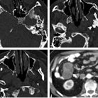
review of skull lesions. Gardner Syndrome (multiple osteomas). Axial head CT images (a–c) show multiple osteomas in the ethmoid air cells (dashed arrows), left ramus of the mandible (arrowhead) and right anterior wall of the maxillary sinus (thin arrow). This patient was also found to have a mesenteric desmoid tumour (thick arrow, d)

Osteoma •
Gardner syndrome - Ganzer Fall bei Radiopaedia

Gardner
syndrome • Gardner syndrome - Ganzer Fall bei Radiopaedia

Gardner
syndrome • Gardner syndrome - Ganzer Fall bei Radiopaedia

Gardner
syndrome • Gardner syndrome - Ganzer Fall bei Radiopaedia

Imaging of
skull vault tumors in adults. Osteomas. Multiple in Gardner syndrome. NECT show mass-like proliferation of normal appearing cortical bone in the frontal (arrow) and cortical and medullary bones in the parieto-occipital (dashed arrow) lesions

Desmoid tumor
in Gardner"s Syndrome presented as acute abdomen. CT scan of the abdomen with contrast media reveals a large-size intra-abdominal mass displacing the adjacent structures. In the same scan a suspicious lesion is identified in the right adrenal. In some rare cases desmoid tumors may co-exist with adrenal or thyroid carcinomas and adrenal adenomas.
Gardner syndrome is one of the polyposis syndromes. It is characterized by:
- familial adenopolyposis
- multiple osteomas: especially of the mandible, skull, and long bones
- epidermal cysts
- fibromatoses
- desmoid tumors of mesentery and anterior abdominal wall
Other abnormalities include:
Pathology
There is an autosomal dominant inheritance in the FAP gene (chromosome 5q) in a majority of patients but with 20% of cases resulting from new mutations. Extracolonic features often precede the diagnosis of colonic polyps.
History and etymology
First described in 1953 by Gardner and Richards .
Siehe auch:
- Osteom
- Lipom
- epidermale Inklusionszyste
- papilläres Schilddrüsenkarzinom
- Leiomyom
- polyposis syndromes
- Desmoid-Tumor - aggressive Fibromatose
- Osteom der Mandibula
- Familiäre adenomatöse Polyposis
- Fibrom
- Gardner's syndrome associated fibromas
- multiple Osteome
- Desmoid-Tumor des Abdomens
- abnormal dentition
- obligate Präkanzerose
und weiter:

 Assoziationen und Differentialdiagnosen zu Gardner-Syndrom:
Desmoid-Tumor
- aggressive Fibromatose


