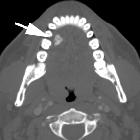
Osteom der Mandibula


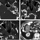
Osteom der Mandibula
Osteom Radiopaedia • CC-by-nc-sa 3.0 • de
Osteomas are benign mature bony growths, seen almost exclusively in bones formed in membrane (e.g. skull).
Terminology
When they arise from bone they may be referred to as a "homoplastic osteoma", and when they arise in soft tissue they may be referred to as a "heteroplastic osteoma".
Clinical presentation
These lesions are benign, slow growing, and usually asymptomatic. They may be incidentally identified as a mass in the skull or mandible, or as the underlying cause of sinusitis or mucocele formation within the paranasal sinuses. When they are multiple, Gardner syndrome should be considered.
They commonly occur in the head and neck, with the most common locations including:
- paranasal sinus osteoma
- ivory osteoma seen most commonly in this location
- skull vault osteoma
- mandibular osteoma
- nasal bones
Pathology
Osteomas are, as the name suggests, osteogenic tumors composed of mature bone. Three histological patterns are recognized :
- also known as eburnated osteoma
- dense bone lacking Haversian system
- also known as osteoma spongiosum
- resembles 'normal' bone, including trabecular bone often with marrow
- a mixture of ivory and mature histology
Radiographic features
The imaging appearance reflects the underlying pathology, with ivory osteomas appearing as very radiodense lesions, similar to the normal cortex, whereas mature osteomas may demonstrate central marrow.
Treatment and prognosis
Osteomas are benign and only require excision if they cause adjacent complications (e.g. mucocele formation) or mass-effect (functional or cosmetic impairment).
Läsionen der Mandibula Radiopaedia • CC-by-nc-sa 3.0 • de
Mandibular lesions are myriad and common. The presence of teeth results in lesions that are specific to the mandible (and maxilla) and a useful classification that defines them as odontogenic or non-odontogenic. While it may often not be possible to make a diagnosis on imaging alone, this classification is helpful to narrow the differential.
Classification
Although a histological classification is probably the most scientifically sound, as radiologists, we are presented with an image, and therefore it is easier to classify lesions according to location in the mandible and their appearance. For a detailed classification of odontogenic tumors, many more than even the keenest neuro/head and neck radiologist can ever remember, please refer to the 2005 WHO histological classification of odontogenic tumors.
Below the lesions are divided into cystic and solid. Cystic should not be confused with lytic as solid radiolucent lesions can also appear lytic (see: radiolucent lesions of the jaw).
Cystic lesions
- periapical cyst (or radicular cyst): common
- dentigerous cyst (or follicular cyst of the mandible): common
- odontogenic keratocyst (OKC): uncommon
- primordial cyst of the mandible
- Stafne cyst (or static bone cavity): uncommon
- solitary bone cyst of the mandible (or traumatic or haemorraghic bone cysts)
- aneursymal bone cyst (ABC): rare in the mandible
- residual cyst of the mandible
Solid lesions
Odontogenic
- benign
- odontoma: most common odontogenic tumor
- ameloblastoma: relatively common
- odontogenic myxoma (looks just like an ameloblastoma): rare
- calcifying epithelial odontogenic tumor (or Pindborg tumor) (looks just like an ameloblastoma): rare
- cementoblastoma: rare
- ameloblastic fibroma
- adenomatoid odontogenic tumor: rare
- condensing osteitis of the mandible
- malignant
- odontogenic carcinoma (e.g. ameloblastic carcinoma): rare
- odontogenic sarcoma: rare
- odontogenic carcinosarcoma: rare
Non-odontogenic
- benign
- idiopathic osteosclerosis
- cemento-ossifying fibroma
- juvenile ossifying fibroma
- exostosis (or torus mandibularis)
- osteoma
- fibrous dysplasia
- Paget disease
- central giant cell lesion/granuloma (giant cell reparative cyst)
- eosinophilic granuloma
- neurofibroma
- schwannoma
- malignant
- squamous cell carcinoma of mandible: common
- osteosarcoma
- Ewing sarcoma
- chondrosarcoma
- metastasis
- multiple myeloma/plasmacytoma
- lymphoma/leukemia
- fibrosarcoma
- leiomyosarcoma
Siehe auch:


 Assoziationen und Differentialdiagnosen zu Osteom der Mandibula: