Paget disease (bone)


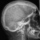


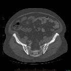


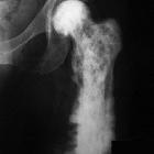


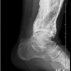
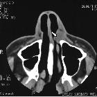


Paget disease of the bone is a common, chronic bone disorder characterized by excessive abnormal bone remodeling. The classically described radiological appearances are expanded bone with a coarsened trabecular pattern. The pelvis, spine, skull, and proximal long bones are most frequently affected.
Epidemiology
It is relatively common and can affect up to 4% of individuals over 40 and up to 11% over the age of 80 . There may be a slight male predilection. Incidence can be considerably higher in the United Kingdom than in other countries. It is also common in Australia, New Zealand, Western Europe, and the United States.
Clinical presentation
The majority (approximately three-quarters) of patients are asymptomatic at the time of diagnosis. Presenting symptoms include:
- localized pain and tenderness
- increased focal temperature due to hyperemia (due to hypervascularity)
- increased bone size: historically changing hat size was a giveaway
- bowing deformities
- kyphosis of the spine
- decreased range of motion
- signs and symptoms relating to complications (see below)
Polyostotic disease is more prevalent than the monostotic type . The most frequent sites of involvement are:
- spine
- pelvis (often asymmetric)
- skull
- proximal long bones
Pathology
The etiology is not entirely known, but it is a disease of osteoclasts. Viral infection (paramyxovirus) in association with genetic susceptibility has been postulated.
There are three classically described stages, which are part of a continuous spectrum :
- early destructive stage (incipient active, lytic): predominated by osteoclastic activity
- intermediate stage (active, mixed): osteoblastic as well as osteoclastic activity
- late stage (inactive, sclerotic/blastic)
These stages correlate well with the imaging findings.
Markers
- elevated serum alkaline phosphatase
- normal calcium and phosphorus levels
- increased urine hydroxyproline
Radiographic features
Signs
There are many Paget disease-related signs, listed here and described in the modality-specific sections below:
- banana fracture
- blade of grass sign
- cotton wool appearance of bone
- ivory vertebra sign
- jigsaw pattern bone or mosaic pattern bone
- Lincoln sign
- Looser zones
- Mickey Mouse sign
- osteoporosis circumscripta
- picture frame vertebra
- Tam o' Shanter sign
Plain radiograph
Plain radiographic features will depend upon the phase of the disease.
The early phase features osteolytic (lucent) regions which are later followed by coarsened trabeculae and bony enlargement. Sclerotic changes occur much later in the disease process.
Additional destructive features may become apparent if malignant transformation occurs.
Skull
- osteoporosis circumscripta: large, well-defined lytic lesions involving the inner aspect of the outer table of the skull (stage one) with a preserved inner table.
- cotton wool appearance: mixed lytic and sclerotic lesions of the skull.
- diploic widening: both inner and outer calvarial tables are involved, with the former usually more extensively affected
- Tam o' Shanter sign: platybasia and basilar invagination with the appearance of the skull falling over the facial bones, like a Tam o' Shanter hat
Spine
- picture frame sign: Paget disease of the spine frequently manifests with cortical thickening and sclerosis encasing the vertebral margins, which gives rise to this appearance on radiographs in mixed-phase disease
- squaring of vertebrae: on lateral radiographs, flattening of the normal concavity of the anterior margin of the vertebral body also adds to the rectangular appearance
- vertical trabecular thickening: coarser than the more delicate pattern seen in intraosseous hemangiomas with which it may be confused
Pelvis
- cortical thickening and sclerosis of the iliopectineal and ischiopubic lines results in the pelvic brim sign and leads to obliteration of Köhler's teardrop
- acetabular protrusion
- enlargement of the pubic rami and ischium
These findings are often asymmetric, and for some reason, are more commonly seen on the right side.
Long bones
- blade of grass or candle flame sign: begins as a subchondral area of lucency with advancing tip of V-shaped osteolysis, extending towards the diaphysis
- in rare cases, the disease is isolated to the diaphysis, most commonly in the tibia, rather than subchondral bone, which can cause diagnostic confusion
- lateral curvature (bowing) of the femur
- anterior curvature of the tibia
MRI
The overall signal characteristics are variable, likely reflecting the natural course of the disease process in different phases.
Several major patterns of involvement have been described:
- dominant signal intensity in Pagetic bone similar to that of fat; most common pattern and probably corresponds to longstanding disease
- relatively low T1 and high T2 signal alteration (also referred as a “speckled” appearance); second most common pattern: probably corresponds to granulation tissue, hypervascularity, and edema seen in early mixed active disease
- low signal intensity on both T1 and T2 images; suggesting the presence of compact bone or fibrous tissue; least common pattern: seen in late sclerotic stage
Fatty marrow signal is usually preserved in all sequences unless there is a complication .
Bone scintigraphy
Tc-99m-MDP is highly sensitive but not specific. It is useful to define the overall extent and distribution of disease.
- marked increased uptake in all phases of the disease, although in the burnt-out sclerotic quiescent phase uptake may be normal
- Mickey Mouse sign: uptake in the vertebral body and posterior elements forming an inverted triangular pattern on posterior planar imaging resembling the Mickey Mouse silhouette ; also known as the "heart" or "clover" sign and "T" or "champagne glass" sign
- Lincoln sign: diffuse mandibular uptake forming a bearded appearance
Treatment and prognosis
Symptomatic patients are treated with bisphosphonates (e.g. alendronate) aiming to reduce the bone turnover, to promote healing of osteolytic lesions and improve bone pain. Analgesics and non-hormonal anti-inflammatory drugs are also prescribed for pain management.
Complications
- osseous weakening resulting in deformity and pathological fractures
- increased risk of osteoarthritis
- hearing loss
- sensorineural hearing loss
- compression of the vestibulocochlear nerve in the internal auditory canal
- loss of bone mineral density of the cochlear capsule
- conductive hearing loss
- fixation of the middle ear ossicles
- sensorineural hearing loss
- neural compression
- cranial nerve paresis may occur
- basilar invagination may occur in advanced cases with hydrocephalus or brainstem compression
- secondary development of tumors (e.g. osteosarcoma: ≈1% of cases, which is often highly resistant to treatment; giant cell tumor in the skull)
- high output congestive cardiac failure
- when bone involvement >15%
- rapid bone formation/resorption can lead to left-to-right shunting and decreased peripheral resistance
- hyperparathyroidism (~10%)
- extramedullary hematopoiesis
History and etymology
Sir James Paget first described it in 1877 in a case report of a patient he had observed over some twenty years .
The condition was initially named by Paget "osteitis deformans", implying an inflammatory etiology. The term "osteodystrophica deformans" is now preferred.
Differential diagnosis
For skull lesions consider
- hyperostosis frontalis interna: thickening of the internal table of the frontal bone
- fibrous dysplasia
- different age group
- fibrous dysplasia usually affects the outer table more prominently
For spinal lesions consider
- vertebral hemangioma: usually finer trabecular markings in hemangioma than Paget related changes
Siehe auch:
- Hyperostosis frontalis interna
- Fibröse Dysplasie
- Morbus Paget der langen Röhrenknochen
- Morbus Paget Becken
- Rahmenwirbel
- Knochenmetastasen
- Ostitis fibrosa cystica
- Osteoporosis circumscripta
- Morbus Paget Szintigraphie
- Grashalm-Zeichen Morbus Paget
- sarkomatöse Entartung bei Morbus Paget des Knochens
- Morbus Paget (Begriffsklärung)
und weiter:
- osteoblastische Knochenmetastasen
- diffuse bony sclerosis (mnemonic)
- Osteopoikilose
- Basiläre Impression
- Coxa vara
- Superscan Szintigraphie
- solitäre lytische Läsion des Schädels
- Protrusio acetabuli
- Thoracic-outlet-Syndrom
- Rugger-Jersey-Wirbelsäule
- vegetable and plant inspired signs
- Biegungsbruch
- Loosersche Umbauzonen
- Morbus Paget der Kalotte
- Osteomalazie
- Insuffizienzfraktur
- Morbus Paget des Femurs
- Leontiasis ossea
- Tibiaverbiegung bei Kindern
- inanimate object inspired signs
- teleangiektatisches Osteosarkom
- Platybasie
- Sandwich-Wirbel
- Hyperostosis Kalotte
- Verbiegung der langen Röhrenknochen
- Morbus Paget der Rippen
- Knochen-in-Knochen-Aspekt
- basilar invagination (mnemonic)
- ivory vertebra sign
- fruit inspired signs
- diffuse skelettale Sklerosierung
- Läsionen des Sakrums
- multiple benign lucent bone lesions (mnemonic)
- vertebral body squaring
- gemischt osteolytisch osteoblastische Knochenmetastasen
- Sklerosierung der Klavikula
- Morbus Paget der Skapula
- very bizarre generalised lesions (mnemonic)
- Bananen-Fraktur
- Morbus Paget des Femurs mit pathologischer Fraktur
- paget disease of a single vertebra
- osteosarcoma of the pelvis in an old woman
- flame sign
- expansive Knochenmetastasen
- Paget's disease of the patella
- Morbus Paget Mittelhandknochen
- Mosaikmuster des Knochens
- Paget disease of the patella
- paget disease - thumb
- Morbus Paget des Sternums
- monostotischer Morbus Paget
- Morbus Paget Humerus

 Assoziationen und Differentialdiagnosen zu Morbus Paget des Knochens:


