Osteoporosis circumscripta


Osteoporosis circumscripta cranii (also known as osteolysis circumscripta) refers to discrete radiolucent regions of the skull on plain radiographs. They are often seen in context of the lytic (incipient-active) phase of Paget disease of the skull, but may be observed in other circumstances as well, e.g. hyperparathyroidism, leontiasis ossea .
Terminology
Over time there has been some debate whether the term should be restricted to non-Pagetoid skull lesions or not . Just like contemporary literature this article uses the term synonymously with the early phase of Paget disease of the skull.
Radiographic features
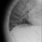
Plain radiograph
Imaging appearances are those of:
- well-defined areas of radiolucency, often large
- most commonly located in frontal and occipital bones
- affecting both inner and outer calvarial tables, with changes in outer table usually more extensive
In patients with simultaneous halisteretic metabolic bone changes, e.g. osteoporosis, identification of above mentioned changes may be challenging.
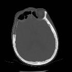
CT
Bony changes are largely identical to those depicted in radiographic findings. Attenuation values of bone marrow space in affected areas are often those of macroscopic fat.
As Paget disease most often presents as a continuum rather than distinct phase, sclerotic areas of the blastic (mixed) phase may more readily be detected. They usually present as cortical and trabecular thickening in a disorganized pattern without bony destruction.
MRI
Cortical changes are more readily appreciated on plain film or CT.
Typical bone marrow changes in the incipient-active to early mixed phase show heterogenous signal on both T1 and T2-weighted imaging :
- T1: decreased signal intensity, isointense to muscle, with interspersed foci of normal signal representing maintained yellow marrow (usable criterion to exclude malignant transformation)
- T2: high intensity changes, heterogeneous and of "speckled" appearance, possibly presenting fibrovascular marrow changes
- C+ (Gd): enhancement, typically of "speckled" appearance, may be present, indicating increased blood flow in the metabolically active sites
Nuclear medicine
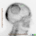
SPECT/CT
Bone-seeking tracers (Tc99m conjugated bisphosphonates, typically MDP) show intense uptake in this early form of Paget disease. Additional sites (polyostotic disease) may be revealed in up to 30% of cases .
In polyostotic disease discrepancy between morphological (CT) and pathophysiological changes (SPECT) especially in early and late stages should not lead to confusion and are well-described in literature .
PET/CT
Non-FDG PET/CT (F18-NaF)
Owing to both higher bone uptake of this tracer combined with superior imaging quality (higher signal-to-noise ratio SNR due to faster blood clearance, higher spatial resolution of PET) compared to SPECT (see above,) intensity of uptake is even higher and polyostotic disease may be even more evident .
FDG PET/CT
Early phase Paget disease of the skull usually lacks FDG-avidity . Encountering a lytic bone lesion (also resembling osteolysis circumscripta) in oncological patients may pose a differential diagnostic dilemma. Finding normal FDG-metabolism may aid in the diagnosis of benign Paget disease and obviate the need for biopsy .
Differential diagnosis
On plain radiograph consider:
- fibrous dysplasia (usually changes more prominent in outer table).
- osteolytic metastases
Siehe auch:
und weiter:


 Assoziationen und Differentialdiagnosen zu Osteoporosis circumscripta: