craniofacial fibrous dysplasia


Craniofacial fibrous dysplasia is one of four types of fibrous dysplasia and is characterized, as the name suggests, by involvement of the skull and facial bones.
For a general discussion of the underlying pathology, refer to the parent article fibrous dysplasia.
Terminology
Although the term leontiasis ossea has been used synonymously with craniofacial fibrous dysplasia, its use is discouraged, as the former has been used to describe a number of entities.
Epidemiology
Young adult patients are most frequently affected. Refer to fibrous dysplasia article for a discussion of epidemiology.
Clinical presentation
Craniofacial involvement may occur both as true craniofacial fibrous dysplasia, considered a form of monostotic fibrous dysplasia (despite multiple cranial bones being affected) that accounts for 10-25% of monostotic cases, or as part of polyostotic fibrous dysplasia. The craniofacial bones are affected in up to 50% of polyostotic cases . Occasionally it is seen in the setting of McCune-Albright syndrome .
Presentation is usually cosmetic or due to mass effect on cranial structures:
- cranial asymmetry
- facial deformity
- nasal stuffiness
- proptosis
- visual impairment/unilateral blindness
Pathology
Refer to fibrous dysplasia article for a discussion of pathology.
Location
The anterior craniofacial bones are more frequently involved than more lateral or posterior portions; sphenoid, frontal, maxillary, ethmoid bones > occipital, temporal bones. Extracranial involvement is rare .
Radiographic features
Similar to fibrous dysplasia elsewhere, affected bones demonstrate a variety of radiographic features ranging from lucency to sclerosis.
Plain radiograph
- blistering/bubbling cystic skull vault lesions
- commonly cross sutures
- sclerotic skull base
- widened diploic space with displacement of outer table; inner table spared (this is in contrast to Paget disease, in which case the inner table is involved)
- obliteration of paranasal sinuses
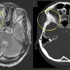
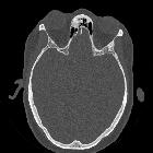
CT
Affected bones are usually expanded with an intact cortex and lose the normal corticomedullary differentiation, being replaced classically by a homogeneous ground glass appearance, although mixed lucencies and sclerosis are also common .
The margin between abnormal and normal bone is often difficult to identify, the two regions blending with each other; however, on occasion, a relatively sharp demarcation may be present . Sometimes the mixed regions of sclerosis and lucency are reminiscent of Paget disease, and are thus referred to as 'pagetoid' .
When the maxilla or mandible are involved, resorption of the roots of teeth is uncommon .
MRI
MRI appearance is variable depending on the degree of lucencies versus sclerosis .
- T1: heterogeneous signal, usually intermediate
- T2: heterogeneous signal, usually low, but may have regions of higher signal
- T1 C+ (Gd): heterogeneous contrast enhancement
Treatment and prognosis
Treatment is reserved for cases where function is being threatened (particularly the airway or vision) and is surgical, although much controversy exists over the best approach (e.g. early vs late intervention, minimal vs radical resection).
As the region affected is often large and involves complex facial anatomy, complete resection is usually not possible. Likewise, reconstruction can be challenging .
Historically, radiotherapy was used to attempt to control growth. This not only was of limited success but encouraged sarcomatous degeneration, and is thus now contraindicated .
Differential diagnosis
General imaging differential considerations include:
- cemento-ossifying fibroma
- histology may be similar; however, trabeculae are rimmed by osteoblasts
- usually more sharply defined
- intraosseous meningioma
- may appear very similar
- usually abuts intracranial compartment
- Paget disease
- predilection for skull vault
- usually spares facial skeleton
- sclerotic metastases
- usually little expansion
- usually has different demographics
Siehe auch:
- Keilbeinmeningeom
- osteoblastische Knochenmetastasen
- Fibröse Dysplasie
- Osteom NNH
- intraossäres Meningeom
- Morbus Paget der Kalotte
- Fibröse Dysplasie im Clivus
- fibröse Dysplasie der Kalotte
- Tumoren am Keilbeinflügel
- cemento-ossifying fibroma
- Fibröse Dysplasie der Schädelbasis
- fibröse Dysplasie am Keilbeinflügel
- fibröse Dysplasie der Mandibula
- kraniofaziale fibröse Dysplasie Nervus opticus
- polyostotic craniofacial fibrous dysplasia in infancy
- Melnick-Needles-Syndrom
- fibrous dysplasia of the temporal bone
- fibrous dysplasia of the orbital roof
und weiter:


 Assoziationen und Differentialdiagnosen zu kraniofaziale fibröse Dysplasie:


