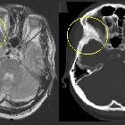
Fibröse Dysplasie der Schädelbasis

de:Fibröse
Dysplasie des rechten de:Jochbeines (im Bild links, markiert). Korrespondierende T2-gewichtete Kernspintomographie (links) und CT (rechts) desselben Patienten Selbsterstellt, Lizenz: Public domain --MBq 16:03, 28. Jun 2006 (CEST)

Radiological
review of skull lesions. Fibrous dysplasia. Axial (a) and coronal (b) head CT images demonstrate an expansile lesion with ground-glass matrix in the right greater wing of the sphenoid (arrows). Axial T1-weighted (c), axial T2-weighted (d) and axial post-contrast T1-weighted (e) images show that the lesion is hypointense with homogeneous enhancement (arrowheads)
Fibröse Dysplasie der Schädelbasis
Siehe auch:

 Assoziationen und Differentialdiagnosen zu Fibröse Dysplasie der Schädelbasis:


