intraosseous meningioma


Intraosseous meningioma, also referred to as primary intraosseous meningioma, is a rare subtype of meningioma that accounts for less than 1% of all osseous tumors. They are the most common type of primary extradural meningiomas .
Terminology
It is important to note that it has been argued by some that this group of meningiomas does not include those intradural meningiomas which present with an intraosseous extension even when the intracranial (non-osseous component) is a minor feature of the mass. For example, en plaque meningiomas often have prominent hyperostosis or bony invasion. Some authors have suggested that this distinction is somewhat arbitrary , and it is certainly true that the terms en plaque meningioma and intraosseous meningioma are often used interchangeably. In the absence of a clearly nodular intracranial component, it is unclear if the distinction can be made on imaging, although en plaque meningiomas are considered far more common.
As a general approach, it is probably reasonable to use the term 'intraosseous' meningioma when there is little if any intracranial extra-osseous disease, and use the term 'en plaque' where there is a definite, albeit sessile, intracranial mass.
Epidemiology
As with meningiomas in general, there is a recognized female predilection.
Clinical presentation
Clinical presentation is usually due to mass effect, the manifestations of which will depend on the location. The calvaria and vertebral column are the most frequent sites . Presentations include:
- palpable or visible bony mass
- proptosis
- cranial nerve / spinal cord compression
- intracranial mass effect/hydrocephalus
Pathology
Thought to occur from trapped arachnoid meningothelial cap cells within cranial sutures during development. However, despite this theory, only a small proportion of intraosseous meningiomas actually occur in association with a skull suture .
Radiographic features
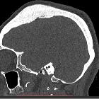
The majority ~65% are osteoblastic while ~35% are osteolytic . Due to this, imaging appearances are often nonspecific.
CT
The commoner osteosclerotic type tends to show diffuse sclerosis with bony expansion.
MRI
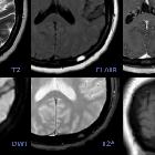
- T1: may show an isointense extra-axial mass component with the expanded bony component being low signal similar to the rest of the skull
- T2: meningioma component is typically isointense to grey matter while a small proportion can be hyperintense
- T1 C+ (Gd): as with conventional meningiomas typically tends to have a uniform avid contrast enhancement
Treatment and prognosis
They are generally benign and slow-growing but there may be a higher proportion of malignant change compared with standard meningiomas . Surgical resection with bone grafting may be performed in symptomatic cases.
Differential diagnosis
For osteoblastic type consider:
- Paget disease: heterogeneous signal, non-enhancing
- craniofacial fibrous dysplasia: tends to be more extensive with more bony remodeling
- osteoma: non-enhancing, more homogeneous contours
- osteosarcoma: irregular contours, heterogeneous signal and enhancement
- osteoblastic metastasis
For osteolytic type consider:
- cranial vault plasmacytoma
- lytic metastasis
Siehe auch:
- Tumoren der Schädelkalotte
- Keilbeinmeningeom
- Osteom Schädelkalotte
- osteoblastische Knochenmetastasen
- Meningeom
- Falxmeningeom
- Meningeom mit Ausdehnung durch die Schädelkalotte
- kraniofaziale fibröse Dysplasie
- Morbus Paget der Kalotte
- fibröse Dysplasie der Kalotte
- En Plaque Meningiom
- osseous meningioma
- intracranial meningiomas
und weiter:

 Assoziationen und Differentialdiagnosen zu intraossäres Meningeom:


