basilar invagination


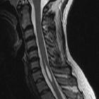
Basilar invagination, also called basilar impression, is a congenital or acquired craniocervical junction abnormality where the tip of the odontoid process projects above the foramen magnum.
Terminology
The terms basilar invagination and basilar impression are often used interchangeably because in both cases there is upwards migration of the upper cervical spine, but they are not synonyms.
Basilar invagination is defined as the congenital upward displacement of vertebral elements into a normal foramen magnum with normal bone.
In contrast, basilar impression is a similar upward displacement of the dens due to, however, acquired softening of bones at the base of skull.
Pathology
It may be congenital or acquired and is often associated with platybasia. There is stenosis of the foramen magnum and compression of the medulla oblongata resulting in neurological symptoms, obstructive hydrocephalus, syringomyelia or even death.
Causes
Common causes can be recalled using the mnemonic PF ROACH. Another approach is to divide them into congenital and acquired causes:
Congenital
- osteogenesis imperfecta
- Klippel-Feil syndrome
- achondroplasia
- Chiari I malformation/Chiari II malformation
- cleidocranial dysostosis
- Schwartz-Jampel syndrome
Acquired
Basilar invagination occurs in around 5-10% of patients with cervical rheumatoid arthritis due to loss of axial supporting structures in the upper cervical spine .
Classification
A classification system has been proposed based on the absence (group I) and presence (group II) of Chiari malformation and can be of use in planning surgical management. Brainstem compression relates to odontoid process indentation in group I, while reduced posterior cranial fossa volume is the cause in group II .
Radiographic features
Five lines have been described and used by most of the radiologists to evaluate basilar invagination on frontal and lateral skull plain radiographs as well as coronal and midsaggital reconstructed CT and MRI images.
This is based on the relative projection of the odontoid process to these lines, which is as follows:
Frontal image
- digastric line: the tip of the odontoid process is normally located 11 to 21 mm below this line
- bimastoid line: the tip of odontoid process projects normally not more than 10 mm above this line
Lateral image
- McRae line: connecting basion and opisthion
- the tip of the odontoid process normally projects below this line, therefore, basilar invagination is diagnosed when the tip crosses this line
- Chamberlain line: connecting the posterior border of hard palate and opisthion
- the tip of the odontoid process projects normally not more than 3 mm above this line
- McGregor line: connecting the posterior edge of the hard palate to the most caudal point of the occipital curve
- the tip of the odontoid process projects normally not more than 5 mm above this line
Siehe auch:
- Morbus Paget des Knochens
- Osteogenesis imperfecta
- Rheumatoide Arthritis
- Chiari-Malformation Typ 1
- Rachitis
- Klippel-Feil-Syndrom
- Kleidokraniale Dysplasie
- Arnold-Chiari-Malformation Typ 2
- Achondroplasie
- primärer Hyperparathyreoidismus
- Syringomyelie
- Osteomalazie
- Verschlusshydrocephalus
- Platybasie
- Chiari-Malformation
- McRae-Linie
- Foramen magnum Stenose bei Achondroplasie
- PF ROACH
und weiter:


 Assoziationen und Differentialdiagnosen zu Basiläre Impression: