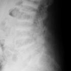
primärer Hyperparathyreoidismus

Lost bones:
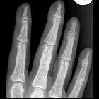
differential diagnosis of acro-osteolysis seen by the pediatric rheumatologist. Frontal radiograph of the left hand in a 14 year old male patient with hyperparathyroidism demonstrates acro-osteolysis (arrowheads), subperiosteal resorption at the middle phalanges (small arrows), brown tumors (large arrows) and diffuse osteopenia
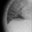
Rugger-Jersey-Wirbelsäule
bei Hyperparathyreoidismus im seitlichen Röntgenbild des Thorax: Grund- und Deckplatten sind deutlich verdichtet.

Brown tumours
(ECR 2016 Case of the Day). Choline PET/CT, axial sections of the pelvis A. CT window B. fusion window.

Generalized
increased bone density in adults • Renal osteodystrophy and brown tumors - Ganzer Fall bei Radiopaedia

Hyperparathyroidism
• Acro-osteolysis from end-stage renal failure - Ganzer Fall bei Radiopaedia
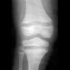
School ager
with renal failure. AP radiograph of the knee shows widening of the distal physis of the femur and the proximal physis of the tibia and fibula and frayed metaphyses in the same areas.The diagnosis was secondary hyperparathyroidism.

Brown tumours
(ECR 2016 Case of the Day). Radiograph of the hand, PA

Brown tumours
(ECR 2016 Case of the Day). Radiograph of the lower limb, AP and lateral.

Brown tumours
(ECR 2016 Case of the Day). CT, axial sections; A and B the lower limb, C and D the lower jaw.

Brown tumours
(ECR 2016 Case of the Day). MRI of the lower limb, coronal sections A. T2-weighted fat-saturated image; B. T1-weighted fat-saturated post contrast image.

Brown tumours
(ECR 2016 Case of the Day). MRI of the lower limb, axial sections; A. T1-weighted image, B. T1-weighted post-contrast image.

Teenager with
seizures and an elevated parathyroid hormone level. Bone windows from an axial CT without contrast of the brain shows salt and pepper lesions diffusely throughout the skull.The diagnosis was primary hyperparathyroidism.
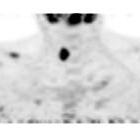
Brown tumours
(ECR 2016 Case of the Day). Choline PET/CT, coronal sections of the neck, PET window.
Hyperparathyroidism
• Rugger-jersey spine - Ganzer Fall bei Radiopaedia

Hyperparathyroidism
• Subperiosteal resorption - Ganzer Fall bei Radiopaedia
Hyperparathyroidism
• Hyperparathyroidism - musculoskeletal manifestations in the hands - Ganzer Fall bei Radiopaedia

Hyperparathyroidism
• Hyperparathyroidism with erosion of dens - Ganzer Fall bei Radiopaedia

Hyperparathyroidism
• Hyperparathyroidism: subperiosteal bone resorption - Ganzer Fall bei Radiopaedia

Hyperparathyroidism
• Renal osteodystrophy - metabolic superscan - Ganzer Fall bei Radiopaedia

Hyperparathyroidism
• Distal clavicular erosions from hyperparathyroidism - Ganzer Fall bei Radiopaedia

Hyperparathyroidism
• Rugger jersey spine - Ganzer Fall bei Radiopaedia

Hyperparathyroidism
• Hyperparathyroidism - Ganzer Fall bei Radiopaedia

Soft tissue
calcification • Hyperparathyroidism - Ganzer Fall bei Radiopaedia
primärer Hyperparathyreoidismus
Siehe auch:
- Weichteilverkalkungen
- Rugger-Jersey-Wirbelsäule
- Renale Osteodystrophie
- Akroosteolyse
- Hyperparathyreoidismus
- Nebenschilddrüsenadenom
- Osteomalazie
- multiple endokrine Neoplasie Typ 1
- subperiostale Knochenresorption
- Ostitis fibrosa cystica
- MEN IIa
- chronische Niereninsuffizienz
- Engel-von-Recklinghausen-Syndrom
- Leitlinie Primärer Hyperparathyreoidismus
- skeletal manifestations of primary hyperparathyroidism
- tertiärer Hyperparathyreoidismus
- selective blood sampling hyperparathyroidism
- superior and inferior rib notching


 Assoziationen und Differentialdiagnosen zu primärer Hyperparathyreoidismus:


