Renale Osteodystrophie

Salt and
pepper sign (skull) • Renal osteodystrophy of the skull vault - Ganzer Fall bei Radiopaedia

Renal
osteodystrophy: A 57-year-old female with end-stage renal disease which results in prominent subchondral resorption resulting in widening of bilateral sacroiliac joints along the iliac surface on AP view of the pelvis (solid arrows). Soft tissue calcifications adjacent to right greater trochanter (dashed arrows), vascular calcification adjacent to the left femoral head (double dashed arrows), and a right femoral dialysis catheter.

Renal
osteodystrophy • Renal osteodystrophy - hand - Ganzer Fall bei Radiopaedia

Brown tumor
• Brown tumor - Ganzer Fall bei Radiopaedia

Brown tumor
• Renal osteodystrophy - Ganzer Fall bei Radiopaedia

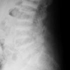
Rugger jersey
spine (hyperparathyroidism) • Renal osteodystrophy - Ganzer Fall bei Radiopaedia

Rugger jersey
spine (hyperparathyroidism) • Renal osteodystrophy - Ganzer Fall bei Radiopaedia

Salt and
pepper sign (skull) • Renal osteodystrophy of the skull vault - Ganzer Fall bei Radiopaedia

Brown tumor
• Brown tumors compressing the spinal cord - Ganzer Fall bei Radiopaedia
Hyperparathyroidism
• Rugger-jersey spine - Ganzer Fall bei Radiopaedia

Calcinosis in
chronic renal disease. Anterior neck and chest SPEC-CT imaging after IV Technetium 99 sestamibi administration shows increased activity of parathyroid glands in addition to normal physiologic activity of the parotid glands, submandibular glands, liver, and myocardium.

Renal
osteodystrophy: AP view of the pelvis (a) in a 26-year-old female with secondary hyperparathyroidism that results in multiple, circumscribed, and expanded lucent areas compatible with brown tumors. Associated fracture deformity of the right femoral head and generalized demineralization.


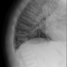
The clothes
maketh the sign. Bone scintigram in a 25-year-old man with renal failure and arthralgia. A frontal planar image (a) and magnified view (b) demonstrates expansion of the manubrium and sternum without manubriosternal joint expansion resulting in a necktie appearance. In addition, a prominent calvarium and faint visualization of the kidneys are also supportive of a diagnosis of renal osteodystrophy

Radiological
review of skull lesions. Renal osteodystrophy and osteopenia. Patient 1: Axial (a) and coronal (b) head CT images depict granular de-ossification with a “pepper pot” appearance (thick arrows) and loss of distinction of the inner and outer tables (dashed arrows) in this patient with renal osteodystrophy. Patient 2: Axial (c) and coronal (d) head CT images show demineralisation of the skull (arrowheads) in this patient with osteopenia. Note the relative preservation of the distinction of the inner and outer tables in osteopenia

Renal
osteodystrophy • Brown tumors - Ganzer Fall bei Radiopaedia

Renal
osteodystrophy • Renal osteodystrophy - Ganzer Fall bei Radiopaedia

Renal
osteodystrophy • Renal osteodystrophy - Ganzer Fall bei Radiopaedia

Renal
osteodystrophy • Renal osteodystrophy with widespread soft tissue calcification - Ganzer Fall bei Radiopaedia

Renal
osteodystrophy • Amyloid arthropathy - Ganzer Fall bei Radiopaedia


Renal
osteodystrophy • Secondary hyperparathyroidism - Ganzer Fall bei Radiopaedia

Hyperparathyroidism
• Subperiosteal resorption - Ganzer Fall bei Radiopaedia

Generalized
increased bone density in adults • Renal osteodystrophy and brown tumors - Ganzer Fall bei Radiopaedia

Generalized
increased bone density in adults • Renal osteodystrophy - Ganzer Fall bei Radiopaedia

Soft tissue
calcification • Hyperparathyroidism - Ganzer Fall bei Radiopaedia

Calcinosis in
chronic renal disease. Plain radiograph of left acromioclavicular calcinosis and osseous erosion of the clavicle, glenoid, and scapula.

Calcinosis in
chronic renal disease. Prominent left femoral artery calcification.

Calcinosis in
chronic renal disease. CT of upper extremity without contrast (coronal) shows calcification of the soft tissues around the acromioclavicular joint with distal clavicle erosion.

Calcinosis in
chronic renal disease. CT upper extremity without contrast (coronal) shows calcification of the soft tissues adjacent to the acromioclavicular joint.

Renal
osteodystrophy • Sacroiliac erosions and subperiosteal resorption from renal osteodystrophy - Ganzer Fall bei Radiopaedia
Renal osteodystrophy (ROD), also known as uremic osteopathy, is a constellation of musculoskeletal abnormalities that occur in patients with chronic renal failure, due to concurrent and superimposed:
- osteomalacia (adults)/rickets (children)
- secondary hyperparathyroidism: abnormal calcium and phosphate metabolism
- bone resorption
- osteosclerosis
- soft tissue and vascular calcifications
- brown tumors
- aluminum intoxication, e.g. if the patient is on dialysis
Radiographic features
Imaging findings are many and varied:
- osteopenia: (often seen early) thinning of cortices and trabeculae
- salt and pepper skull
- demineralization: usually subperiosteal, however, it may also involve joint margins, endosteal, subchondral, subligamentous areas, cortical bone, or trabeculae
- subperiosteal resorption: characteristic subperiosteal resorption may be seen on radial aspects of middle phalanges of index and long fingers
- bone sclerosis
- diffuse bony sclerosis
- rugger jersey spine: sclerosis of the vertebral body endplates
- soft tissue calcification
- amyloid deposition: erosion in and around joint
- insufficiency fractures
- Looser zone
- brown tumors
Differential diagnosis
General imaging differential considerations include
- osteomalacia
- rheumatoid arthritis
- seronegative spondyloarthropathies
- neoplasms: multiple myeloma, metastases; brown tumors can mimic primary malignant tumor of bone; amyloid deposition may mimic PVNS or synovial chondromatosis
- osteomyelitis
- occult marrow abnormalities
Siehe auch:
- synoviale Osteochondromatose
- Multiples Myelom
- primärer Hyperparathyreoidismus
- Pigmentierte villonoduläre Synovialitis
- Osteomalazie
- subperiostale Knochenresorption
- Ostitis fibrosa cystica
- osteomesopyknosis
- Osteodystrophy
- Amyloidarthropathie
- Metastasen
- seronegative Spondyloarthropathie
und weiter:
- diffus hypointenses Knochenmarksignal in T1
- diffuse bony sclerosis (mnemonic)
- Superscan Szintigraphie
- Riesenzelltumor
- dichte metaphysäre Bänder
- Riesenzelltumor des Knochens
- Calciphylaxie
- Gefäßverkalkungen
- renale Osteodystrophie Schädelkalotte
- diffuse skelettale Sklerosierung
- Osteoklastom
- generalized increased bone density in paediatrics
- polyostotic bone lesions in adults
- Rugger Jersey Spine Osteoporose


 Assoziationen und Differentialdiagnosen zu Renale Osteodystrophie:


seronegative
Spondyloarthropathie