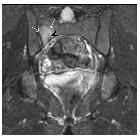
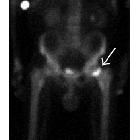
insufficiency fracture


 nicht verwechseln mit: Ermüdungsfraktur
Insufficiency fractures are a type of stress fracture, which are the result of normal stresses on abnormal bone. Looser zones are also a type of insufficiency fracture. They should not be confused with fatigue fractures which are due to abnormal stresses on normal bone, or with pathological fractures, the result of diseased, weakened bone due to focal pathology such as tumors (both malignant and benign).
Epidemiology
In general, they are seen in the elderly, more frequently in women .
Pathology
They are most often seen in the setting of osteoporosis, although any process which weakens bone is a risk factor. Long-term bisphosphonate use has also been associated with insufficiency fractures .
Etiology
Osteoporosis is the most common cause of insufficiency fractures, although there are many more :
- disrupted bone mineral homeostasis: osteoporosis, hyperparathyroidism, diabetes mellitus, osteomalacia
- bone remodeling: Paget disease, osteopetrosis
- collagen formation: Marfan syndrome, fibrous dysplasia
- medications: glucocorticoids, chemotherapy
- radiation therapy
Location
- vertebral (crush or wedge) fractures: very common
- marrow edema is limited to the vertebral body; extension of abnormal signal into the pedicles suggests an underlying lesion
- sacrum: Honda sign
- neck of femur
- proximal third femur (see article: bisphosphonate-related proximal femur fractures)
- pubic rami
- sternum
- fibula
- tibia
Radiographic features
Early diagnosis is best made with bone scan or MRI, as plain films may initially appear normal.
Plain radiograph
- initially normal
- periosteal reaction progressing to callus formation in diaphyseal fractures
- linear sclerosis and cortical thickening more frequent in metaphyseal and epiphyseal fractures
MRI
MRI is as sensitive as bone scanning, with the added benefit of higher specificity, both in isolating the exact anatomic location and in distinguishing fractures from tumors or infection.
- T1: low marrow signal
- T2: high marrow signal with extension into adjacent soft tissues
- C+ (Gd): enhancement can be intense
Nuclear medicine
On bone scan, there is increased activity at the site of the fracture.
Treatment and prognosis
Treatment depends on the location and whether the fracture is complete or incomplete. Options, therefore, include:
- conservative management
- plaster cast
- internal fixation
- vertebroplasty
- kyphoplasty
Treatment of the underlying cause of bone weakness is also essential.
Siehe auch:
- Okkulte Fraktur
- Morbus Paget des Knochens
- pathologische Fraktur
- Stressfraktur
- Osteoporose
- Loosersche Umbauzonen
- Osteomalazie
- Vertebroplastie
- Ermüdungsbruch
- subchondrale Insuffizienzfraktur
- Subchondroplasty
- Insuffizienzfraktur des Sakrums
und weiter:

 Assoziationen und Differentialdiagnosen zu Insuffizienzfraktur:


