renale Osteodystrophie Schädelkalotte

Radiological
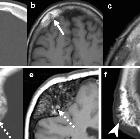
review of skull lesions. Renal osteodystrophy and osteopenia. Patient 1: Axial (a) and coronal (b) head CT images depict granular de-ossification with a “pepper pot” appearance (thick arrows) and loss of distinction of the inner and outer tables (dashed arrows) in this patient with renal osteodystrophy. Patient 2: Axial (c) and coronal (d) head CT images show demineralisation of the skull (arrowheads) in this patient with osteopenia. Note the relative preservation of the distinction of the inner and outer tables in osteopenia

Imaging of
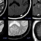
skull vault tumors in adults. Miscellany. Paget disease (a, b) CT (a) and X-ray (b): Characteristic mixed bone lysis and sclerosis, cortical bone thickening, and expansion. Osteoporosis circumscripta cranii CT (c, d): large geographic radiolucent areas involving medullar and cortical bone in frontal and occipital regions (arrows). Amyloidoma (e, f) CT (e) and T2WI (f): Giant heterogeneous mass with marked T2 hypointensity and calcifications. Renal osteodystrophy CT (g): shows characteristic “salt-and-pepper pattern.” Brown tumor (h, i): unspecific well-defined, cystic appearance (arrows). Thalassemia CT (j): Diffuse diploic widening and “hair-on-end” appearance (arrow) with characteristic occipital bone preservation (arrowhead). Bone sarcoidosis (k, l) CT (k) and T2WI (l): Mixed predominantly lytic multiple lesions with lace-like internal pattern of calcification and T2 hypointensity (dashed arrows). Osteitis CT (m, n): Bone focal lysis and erosions of osteitis (arrows) contiguous to a frontal sinusitis complicated with intracranial laminar abscess (dashed arrow)
renale Osteodystrophie Schädelkalotte
Renale Osteodystrophie Radiopaedia • CC-by-nc-sa 3.0 • de
Renal osteodystrophy (ROD), also known as uremic osteopathy, is a constellation of musculoskeletal abnormalities that occur in patients with chronic renal failure, due to concurrent and superimposed:
- osteomalacia (adults)/rickets (children)
- secondary hyperparathyroidism: abnormal calcium and phosphate metabolism
- bone resorption
- osteosclerosis
- soft tissue and vascular calcifications
- brown tumors
- aluminum intoxication, e.g. if the patient is on dialysis
Radiographic features
Imaging findings are many and varied:
- osteopenia: (often seen early) thinning of cortices and trabeculae
- salt and pepper skull
- demineralization: usually subperiosteal, however, it may also involve joint margins, endosteal, subchondral, subligamentous areas, cortical bone, or trabeculae
- subperiosteal resorption: characteristic subperiosteal resorption may be seen on radial aspects of middle phalanges of index and long fingers
- bone sclerosis
- diffuse bony sclerosis
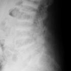
- rugger jersey spine: sclerosis of the vertebral body endplates
- soft tissue calcification
- amyloid deposition: erosion in and around joint
- insufficiency fractures
- Looser zone
- brown tumors
Differential diagnosis
General imaging differential considerations include
- osteomalacia
- rheumatoid arthritis
- seronegative spondyloarthropathies
- neoplasms: multiple myeloma, metastases; brown tumors can mimic primary malignant tumor of bone; amyloid deposition may mimic PVNS or synovial chondromatosis
- osteomyelitis
- occult marrow abnormalities
Siehe auch:


 Assoziationen und Differentialdiagnosen zu renale Osteodystrophie Schädelkalotte: