Chiari I Malformation


 nicht verwechseln mit: benigner Tiefstand der Kleinhirntonsillen / sekundärer Tiefstand der Kleinhirntonsillen
Chiari I malformation is the most common variant of the Chiari malformations and is characterized by a caudal descent of the cerebellar tonsils (and brainstem in its subtype, Chiari 1.5) through the foramen magnum. Symptoms are proportional to the degree of descent. MRI is the imaging modality of choice. Treatment with posterior decompression is usually reserved for symptomatic patients or those with a syrinx.
Epidemiology
Chiari I malformations are more frequently encountered in females .
Clinical presentation
Unlike Chiari II, III, and IV malformations, Chiari I malformations often remain asymptomatic until adulthood.
If present, symptoms may include a headache and those associated with brainstem compression, syringomyelia, or scoliosis.
The likelihood of becoming symptomatic is proportional to the degree of descent of the tonsils. In one study, all patients with greater than 12 mm of descent were symptomatic, whereas approximately 30% of those whose descent measured between 5 and 10 mm remained asymptomatic .
Pathology
The Chiari I malformation is characterized by an inferior position of the cerebellar tonsils relative to the foramen magnum. This is believed to be due to a mismatch between the size and content of the posterior fossa.
Four groups of Chiari I patients can be distinguished, according to different pathogeneses :
Chiari I malformations need to be distinguished from low-lying tonsils (benign tonsillar ectopia) which is an asymptomatic and incidental finding in normal individuals, whereby the tonsils protrude through the foramen magnum by no more than 3-5 mm .
The terminology of caudally displaced tonsils is discussed in the article on cerebellar tonsillar ectopia.
Associations
Although Chiari I malformations are often isolated abnormalities, the following findings may be seen in association:
- cervical cord syrinx in ~35% (range 20-56%): more common in symptomatic patients
- hydrocephalus in up to 30% of cases
- thought to result from abnormal CSF flow dynamics through the central canal of the cord and around the medulla
- skeletal anomalies in ~35% (range 23-45%) :
Radiographic features
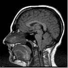
Although historically visible on myelography, cross-sectional imaging (especially MRI) is needed to diagnose accurately and assess for Chiari I malformations. In either case, the diagnosis is made by measuring the cerebellar tonsillar position (TP).
CT
With modern volumetric scanning and high quality sagittal reformats relatively good views of the foramen magnum and tonsils can be achieved although the intrinsic lack of contrast (compared to MRI) makes accurate assessment difficult. More frequently the diagnosis is suspected on axial images where the medulla is embraced by the tonsils and little if any CSF is present. This is referred to as a crowded foramen magnum.
MRI
MRI is the imaging modality of choice. On sagittal imaging, the best plane for assessing for the presence of Chiari I malformations, the tonsils are pointed, rather than rounded and referred to as peg-like. The sulci are vertically oriented, forming so-called sergeant stripes. Axial images through the foramen show crowding of the medulla by the tonsils.
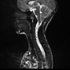
Spinal cord MRI should be recommended as a syrinx may be seen if progressively enlarging or associated with symptoms may be an indication for surgical intervention .
CSF flow studies may also be useful to assess the flow surrounding the cervicomedullary junction.
Importantly, features of intracranial hypertension should be sought to ensure that cerebellar tonsillar ectopia is not secondary to raised intracranial pressure (and therefore not a Chiari I malformation) .
Treatment and prognosis
Chiari I malformations can be divided into three stages (although not frequently used in day to day practice):
- I: asymptomatic
- II: brainstem compression
- III: syrinx
Treatment is usually reserved only for symptomatic patients or those with a syrinx. It consists of decompressing the posterior fossa, by removing part of the occipital bone, and posterior arch of C1 as well as performing a duroplasty.
History and etymology
It was first described in 1891 by Hans Chiari (1851-1916), Austrian pathologist, based on the autopsies of children.
Differential diagnosis
Imaging differential considerations include:
- incidental tonsillar ectopia: <5 mm
- Chiari 1.5 malformation (sometimes considered a variant of Chiari I malformation )
- Chiari II malformation
- acquired tonsillar ectopia
Distinguishing Chiari I malformation for pseudotumor cerebri is particularly important as treatment with a posterior fossa decompression can result in poor outcomes . Examination for features of intracranial hypertension is therefore crucial.
Siehe auch:
- Basiläre Impression
- Arnold-Chiari-Malformation Typ 2
- Myelomeningozele
- Tiefstand der Kleinhirntonsillen
- Chiari-Malformation
- Erweiterungsplastik Foramen magnum
- benigner Tiefstand der Kleinhirntonsillen
- Chiari-Malformation Typ 3
- pädiatrische Erkrankungen des Liquorsystems
- sekundärer Tiefstand der Kleinhirntonsillen
- Chiari-Malformation Typ 4
und weiter:
- Syrinx
- Liquorunterdrucksyndrom
- neuroradiologisches Curriculum
- Syringomyelie
- Einklemmung Foramen magnum
- Crouzon-Syndrom
- basilar invagination (mnemonic)
- Chiari 1.5 malformation
- hydromyelia due to Chiari I malformation
- Laminektomie Atlas
- Chiari-Malformation Typ 1 mit Syrinx
- chiari I malformation with syrinx
- peg like tonsils
- sergeant stripes


 Assoziationen und Differentialdiagnosen zu Chiari-Malformation Typ 1:


