Cerebellum


The cerebellum, meaning "the little brain", sits at the base of the brain in the posterior cranial fossa below the tentorium and behind the brainstem.
Gross anatomy
The cerebellum has the following features:
- three surfaces: anterior (petrosal), superior (tentorial), inferior (suboccipital)
- three fissures: primary (tentorial), horizontal (petrosal), prebiventral/prepyramidal (suboccipital)
- two hemispheres
- single median vermis
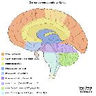
Vermis
The vermis is divided into nine lobules: (in a clockwise rotation, looking at the patient sagittally from his left), and separated into groups by fissures:
- lingula
- central lobule
- culmen: primary (tentorial) fissure
- declive
- folium: horizontal (petrosal) fissure
- tuber: prebiventral/prepyramidal (suboccipital) fissure
- pyramid
- uvula
- nodulus
The subdivisions of the cerebellar vermis can be remembered by this mnemonic.
Cerebellar hemisphere
The cerebellar folia run parallel to the calvaria in an onion-like configuration.
Each of the nine vermis lobules is associated in both sides with two cerebellar hemisphere lobules and therefore the cerebellum has 18 cerebellar hemisphere lobules:
- wing of lingula (lingula)
- wing of central lobule (central lobule)
- quadrangular lobule (culmen): primary (tentorial) fissure
- simple lobule (declive)
- superior semilunar lobule (folium): horizontal (petrosal) fissure
- inferior semilunar lobule (tuber): prebiventral/prepyramidal (suboccipital) fissure
- biventral lobule (pyramid)
- tonsil (uvula)
- flocculus (nodulus)
Connections to brainstem
- to the midbrain via the superior cerebellar peduncles (brachia conjunctiva)
- to the pons via the middle cerebellar peduncles (brachia pontis)
- to the medulla via the inferior cerebellar peduncles (restiform bodies)
CSF cisterns
Arterial supply
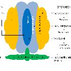
The cerebellum is supplied by three bilateral arteries from the vertebrobasilar system:
Superior cerebellar (SCA)
The SCA supplies:
- whole superior surface of the cerebellar hemispheres down to the great horizontal fissure
- the superior vermis
- dentate nucleus
- most of the cerebellar white matter
- superior cerebellar peduncle
- middle cerebellar peduncle
Anterior inferior cerebellar (AICA)
The AICA has a variable vascular territory depending on the size of the PICA (see AICA-PICA dominance) but usually supplies:
- middle cerebellar peduncle
- inferolateral portion of the pons
- flocculus
- anteroinferior surface of the cerebellum
Posterior inferior cerebellar (PICA)
The PICA has a variable vascular territory depending on the size of the AICA (see AICA-PICA dominance), but usually supplies:
- posteroinferior cerebellar hemispheres (up to the great horizontal fissure)
- inferior portion of the vermis
- inferior cerebellar peduncle
It divides into lateral and medial branches that supply the inferior portion of the vermis and cerebellar hemispheres respectively.
There are some variations in the PICA:
- 18% arise extracranially, inferior to the foramen magnum
- 10% arise from the basilar rather than vertebral artery
- 2% bilaterally absent
- occasionally loops around the cerebellar tonsil
- occasionally a small vertebral artery will terminate into a common PICA/AICA trunk
Venous drainage
There are three major groups of bilateral veins that drain the cerebellum along with other structures of the posterior cranial fossa. Not unsurprisingly there is significant variation between patients and also between the left and right in the same patient.
The three posterior fossa venous groups are:
- superior (Galenic)
- drains the midbrain, upper pons, superior vermis and the superior surface of the cerebellar hemispheres
- drains into the vein of Galen and has three named veins
- precentral cerebellar vein
- superior vermian vein
- anterior pontomesencephalic vein
- anterior (petrosal)
- drains the lateral pons, medulla and the anterior surface of the cerebellar hemispheres
- drains into the inferior petrosal sinus and has a petrosal vein
- posterior (tentorial)
- drains the inferior vermis, tonsils and the posteroinferior surface of the cerebellar hemispheres
- drains into the transverse sinus and has an inferior vermian vein
Variant anatomy
- cerebellar agenesis
- tonsillar ectopia: asymptomatic protrusion of the tonsils through the foramen magnum by no more than 3-5 mm
Related pathology
- neoplastic
- inflammatory
- degenerative
- diffuse cerebellar atrophy
- crossed cerebellar atrophy
- congenital disease
- others
Siehe auch:
- Cisterna magna
- Arteria cerebelli superior
- Arteria cerebelli inferior posterior (PICA)
- cerebellopontine angle
- Arteria cerebelli anterior inferior
- Flocculus
- posterior inferior cerebellar
- anterior inferior cerebellar
und weiter:
- Cisterna cerebellomedullaris
- Circulus Willisi
- Arteria basilaris
- Cisterna quadrigeminalis
- neuroradiologisches Curriculum
- Anatomie Hirnstamm
- posterior cerebral circulation
- vierter Ventrikel
- ambient cistern
- inferior petrosal sinus
- brain development
- extrapyramidal system
- McLeod phenomenon
- aqueduct of Sylvius
- Rosettenbildender glioneuronaler Tumor
- posterior fossa astrocytoma
- rosette-forming glioneuronal tumours
- tuberkulöse Meningitis
- Thrombose Arteria vertebralis
- Anatomie Cerebrum


 Assoziationen und Differentialdiagnosen zu Cerebellum:


